Pediatric patients (0–18 years) with congenital hearing loss
Care Setting
Tertiary care hospital imaging and pediatric ENT services
Key Highlights
Approximately 20% of congenital sensorineural hearing loss cases have radiologically detectable inner ear malformations or nerve abnormalities.
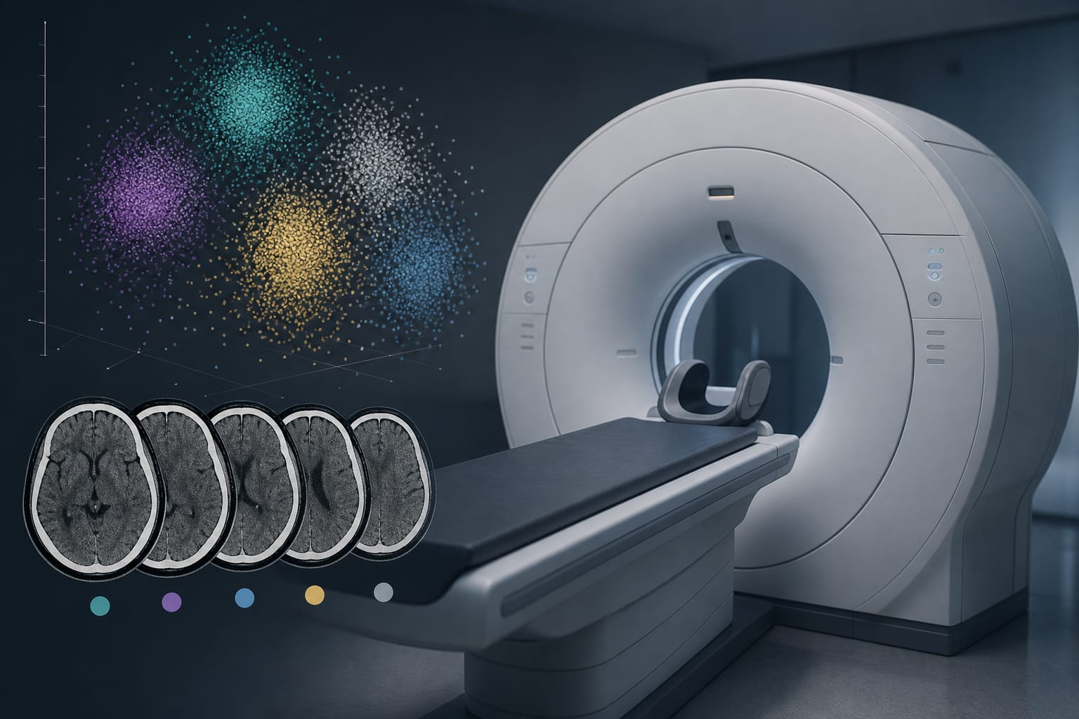
CT and MRI are complementary imaging modalities essential for evaluating osseous and nerve structures in congenital hearing loss.
Sennaroglu’s 2017 classification is the most widely accepted system for categorizing inner ear malformations and guiding management.
Guideline-Based Recommendations
Diagnosis
Use universal newborn hearing screening with two-stage automated auditory brainstem response protocol.
Employ CT and MRI imaging to detect and classify inner ear malformations and nerve abnormalities.
Classify malformations according to Sennaroglu’s 2017 classification for standardized diagnosis.
Management
Early detection of inner ear malformations is critical for timely intervention and rehabilitation.
Audiological and radiological findings guide treatment decisions between cochlear implantation and auditory brainstem implantation.
Monitoring & Follow-up
Baseline audiometry should be performed using age-appropriate methods: VRA (6–18 months), play audiometry (18 months–5/6 years), and pure tone audiometry (≥6–7 years).
Hearing thresholds should be averaged at 0.5, 1, 2, and 4 kHz frequencies to classify hearing impairment severity.
Risks
Delayed diagnosis may lead to missed opportunities for early intervention and poorer hearing outcomes.
Misclassification of malformations may affect treatment planning and prognosis.
Patient & Prescribing Data
Pediatric patients with congenital sensorineural hearing loss and radiologically confirmed inner ear malformations
Audiological and imaging findings are essential to tailor interventions such as cochlear or auditory brainstem implantation.
Clinical Best Practices
Implement universal newborn hearing screening programs with follow-up diagnostic testing for positive screens.
Use combined CT and MRI imaging to comprehensively evaluate inner ear and nerve structures.
Apply standardized classification systems (Sennaroglu 2017) to guide diagnosis and management.
Perform age-appropriate audiological assessments to monitor hearing thresholds and impairment severity.
Conduct multidisciplinary evaluation including radiologists, audiologists, and ENT specialists for optimal care planning.