Choroidal structural and perfusion characteristics across refractive groups in children
-
By
-
Yu Liu
-
Getu Tao
-
Yifan Zhao
-
Mengyao Ma
-
Shuang Feng
-
Min Qin
-
Xiuli Bao
-
June 10, 2026
-
Clinical Scorecard: Choroidal Structure and Blood Flow Variations in Pediatric Refractive Error Categories
At a Glance
| Category | Detail |
|---|
| Condition | |
| Key Mechanisms | Choroidal structural changes and perfusion variations associated with refractive error. |
| Target Population | |
| Care Setting | |
Key Highlights
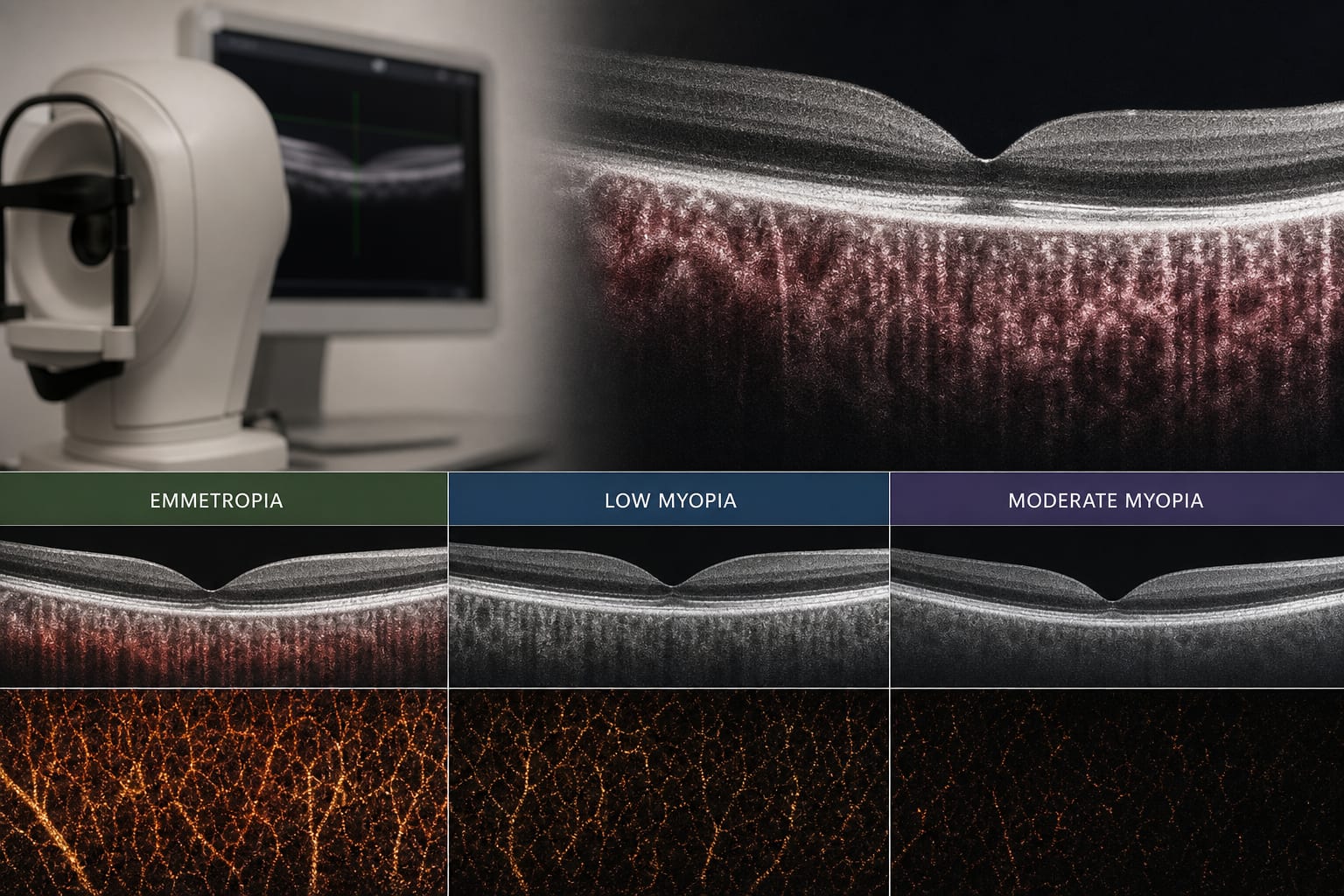
- Choroidal thickness decreases with increasing myopia severity.
- Haller's layer thinning is the primary driver of choroidal structural changes.
- No significant differences in OCTA-derived perfusion parameters between groups.
- OCT-derived structural parameters correlate positively with spherical equivalent refraction (SER) and negatively with axial length (AL).
- Limited associations observed between OCTA-derived parameters and SER or AL.
Guideline-Based Recommendations
Diagnosis
- Classify children into emmetropia, low myopia, and moderate myopia based on cycloplegic SER.
Management
- Monitor choroidal structural changes in children with myopia.
Monitoring & Follow-up
- Use EDI-OCT and OCTA for evaluating choroidal thickness and perfusion.
Risks
- Increased risk of myopia-related ocular complications with longer axial length.
Patient & Prescribing Data
Children aged 8–14 years with varying degrees of myopia.
Clinical Best Practices
- Utilize EDI-OCT for assessing choroidal thickness in pediatric patients.
- Consider both structural and perfusion metrics for comprehensive evaluation.
Related Resources & Content