Impact of spread through air spaces (STAS) and lymphovascular invasion (LVI) on prognosis in NSCLC: a comprehensive pathological evaluation - Scorecard - MDSpire
Advertisement
Impact of spread through air spaces (STAS) and lymphovascular invasion (LVI) on prognosis in NSCLC: a comprehensive pathological evaluation
Clinical Scorecard: Influence of Air Space Spread (STAS) and Lymphovascular Invasion (LVI) on Prognosis in Non-Small Cell Lung Cancer: An In-Depth Pathological Analysis
At a Glance
Category
Detail
Condition
Non-Small Cell Lung Cancer (NSCLC)
Key Mechanisms
Spread Through Air Spaces (STAS) and Lymphovascular Invasion (LVI) as independent pathological features influencing recurrence and survival
Target Population
Patients undergoing surgery for NSCLC including squamous cell carcinoma, adenocarcinoma, and adenosquamous carcinoma
Care Setting
Surgical oncology and pathology departments in tertiary care hospitals
Key Highlights
STAS is defined by malignant cells in alveolar spaces adjacent to the tumor and is associated with poor prognosis and increased recurrence risk, especially in limited resections.
LVI indicates tumor presence in endothelial-lined lymphatic or blood vessels and correlates with higher nodal spread and distant metastasis risk.
TNM staging does not currently include STAS or LVI, although these factors independently affect prognosis and may guide adjuvant therapy decisions.
Guideline-Based Recommendations
Diagnosis
Evaluate STAS by identifying micropapillary clusters, solid nests, or single tumor cells beyond the tumor edge in alveolar spaces using histopathology and immunohistochemistry.
Identify LVI by detecting tumor cells within endothelial-lined lymphatic or vascular channels adjacent to the tumor.
Exclude artifacts such as mechanical floaters or fragmented tumor clusters to avoid misdiagnosis of STAS.
Management
Consider adjuvant chemotherapy for patients with LVI as per NCCN guidelines due to high-risk status.
Incorporate STAS status in surgical planning, especially when considering limited resections for early-stage adenocarcinoma.
Monitoring & Follow-up
Monitor for local recurrence within 2 years post-surgery in adjacent lung tissue and regional lymph nodes, particularly in patients with STAS and/or LVI.
Use Kaplan–Meier survival analysis and Cox regression models to assess recurrence-free and overall survival in follow-up.
Risks
STAS increases risk of tumor recurrence and poorer overall survival.
LVI is associated with higher likelihood of nodal metastasis and distant organ spread, worsening prognosis.
Patient & Prescribing Data
Patients with surgically resected NSCLC, predominantly male, mean age ~60 years, including early and advanced stages.
Adjuvant chemotherapy is recommended for patients with LVI due to increased metastatic risk; STAS presence may influence surgical approach and postoperative management.
Clinical Best Practices
Perform thorough histopathological examination of resected lung specimens to identify STAS and LVI using standardized criteria and immunohistochemical stains.
Exclude artifacts rigorously to prevent false-positive STAS diagnosis.
Integrate STAS and LVI findings with TNM staging to refine prognosis and tailor adjuvant therapy decisions.
Ensure multidisciplinary collaboration between surgeons, pathologists, and oncologists for optimal patient management.
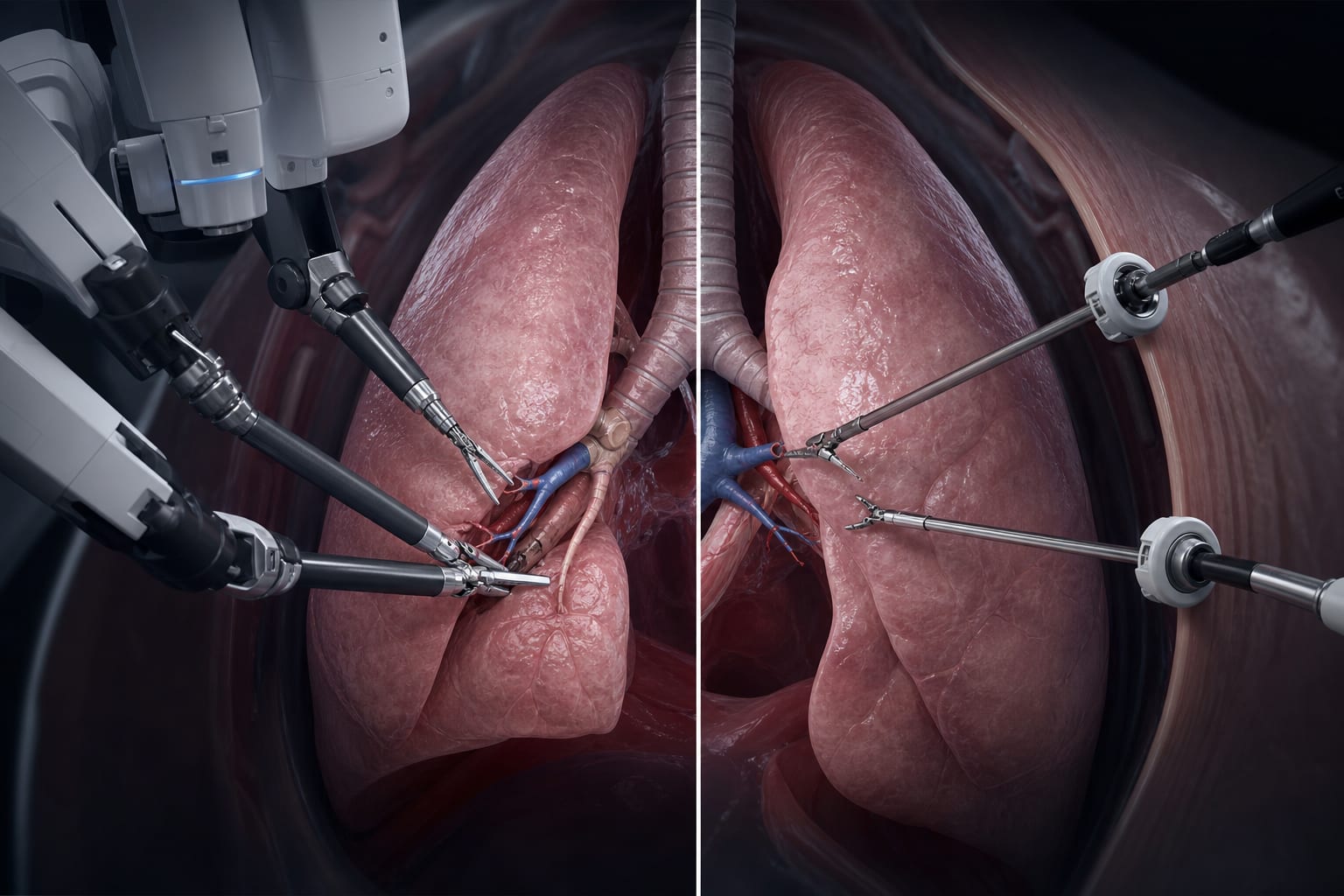
At Morristown and Overlook Medical Centers, robotic bronchoscopy and multidisciplinary collaboration are redefining what’s possible for patients with suspected lung cancer.