Subcortical language localization using sign language and awake craniotomy for dominant posterior temporal glioma resection in a hearing-impaired patient - Scorecard - MDSpire
Advertisement
Subcortical language localization using sign language and awake craniotomy for dominant posterior temporal glioma resection in a hearing-impaired patient
Clinical Scorecard: Localization of Language Functions in Subcortical Areas During Awake Craniotomy for Resection of Dominant Posterior Temporal Glioma in a Patient with Hearing Impairment
At a Glance
Category
Detail
Condition
Intrinsic brain tumor (lower-grade glioma) involving eloquent language areas in the dominant posterior temporal lobe
Key Mechanisms
Intraoperative direct cortical and subcortical stimulation (DCS) during awake craniotomy to map language functions in both spoken and sign language modalities
Target Population
Patients with dominant hemisphere gliomas affecting language areas, including those with hearing impairment and bilingual communication (spoken and sign language)
Care Setting
Neurosurgical operating room with awake craniotomy and intraoperative neurophysiological monitoring
Key Highlights
First reported case of awake language mapping testing both sign language and spoken language modalities in a post-lingual deaf patient.
Preoperative functional MRI and diffusion tensor imaging (DTI) used to localize language areas and critical white matter tracts (arcuate fasciculus and inferior fronto-occipital fasciculus) near the tumor.
Subcortical stimulation elicited phonemic paraphasias, guiding the extent of tumor resection while preserving language function.
Guideline-Based Recommendations
Diagnosis
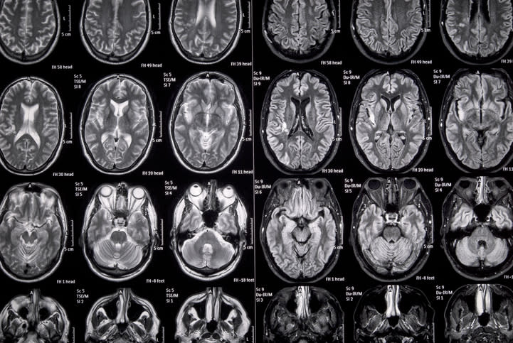
Use MRI with T1, T2, and FLAIR sequences to characterize tumor and involvement of eloquent cortex.
Perform preoperative task-based functional MRI to lateralize language and identify activation near lesions.
Use diffusion tensor imaging tractography to delineate critical language white matter pathways.
Management
Consider awake craniotomy with intraoperative direct cortical and subcortical stimulation for maximal safe resection of tumors near language areas.
Test both spoken and sign language modalities intraoperatively when applicable, with collaboration from neuropsychology and sign language interpreters.
Use neuromonitoring and neuronavigation integrated with preoperative imaging to guide resection boundaries.
Monitoring & Follow-up
Monitor language function intraoperatively using direct stimulation and naming tasks with visual stimuli.
Observe for language errors such as phonemic paraphasias to identify functional language pathways during resection.
Risks
Risk of postoperative language deficits if eloquent cortex or subcortical language tracts are damaged.
Potential challenges in intraoperative language testing in hearing-impaired patients requiring tailored approaches.
Patient & Prescribing Data
Post-lingual deaf patient with profound sensorineural hearing loss and bilingual communication in American Sign Language and English.
Awake craniotomy with intraoperative mapping can be successfully performed in hearing-impaired patients using both spoken and sign language stimuli to preserve language function.
Clinical Best Practices
Integrate preoperative fMRI and DTI tractography to plan surgical approach and anticipate language pathway locations.
Use awake craniotomy with direct cortical and subcortical stimulation to maximize tumor resection while preserving language.
Collaborate with neuropsychology and sign language interpreters for intraoperative testing in bilingual and hearing-impaired patients.
Employ naming tasks with visual stimuli for both spoken and sign language during mapping.
Use intraoperative neuro-navigation combined with stimulation findings to define safe resection margins.
These 10 states make it more practical for physicians to participate in hospital ownership by aligning statutory structure, corporate practice of medicine rules, and population trends.