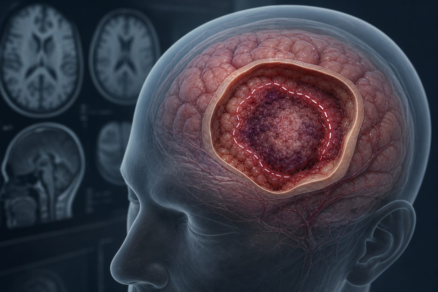
Aneurysm formation influenced by vascular angulation, wall shear stress (WSS), oscillatory shear index (OSI), and hemodynamic factors at arterial bifurcations
Target Population
Adults (≥18 years) with unruptured intracranial aneurysms confirmed by angiography
Care Setting
Neurosurgical centers with neurovascular multidisciplinary teams and advanced imaging capabilities (MRA, CTA, DSA)
Key Highlights
MIAs are bilateral aneurysms on corresponding arteries, often located at high turbulent flow sites like MCA and ICA terminus.
Aneurysm multiplicity, including MIAs, is associated with increased rupture risk compared to singular aneurysms.
Risk stratification models incorporate aneurysm morphology (size, aspect ratio, size ratio) and patient factors to guide elective treatment decisions.
Guideline-Based Recommendations
Diagnosis
Confirm unruptured intracranial aneurysms via angiographic imaging (CTA, MRA, or DSA).
Classify aneurysms into singular, asymmetrical multiple, or mirror intracranial aneurysms based on anatomical criteria.
Exclude mycotic, vasculitic, AVM-associated, extradural, previously treated aneurysms, and lesions too small to confirm as aneurysms.
Management
Individualize treatment decisions by weighing rupture risk against procedural risks.
Consider elective intervention for high-risk aneurysms identified by morphology and multiplicity.
Monitor MIAs closely due to their anatomical clustering at high-stress bifurcations and potential increased rupture hazard.
Monitoring & Follow-up
Perform longitudinal follow-up with repeat angiographic imaging to assess aneurysm growth and morphological changes.
Document symptoms at diagnosis and during follow-up to detect clinical changes.
Use multidisciplinary team review to integrate imaging and clinical data for ongoing risk assessment.
Risks
Aneurysmal subarachnoid hemorrhage carries approximately 50% mortality at 1 month.
Multiplicity of aneurysms, including MIAs, increases rupture risk compared to singular aneurysms.
Treatment risks must be balanced against rupture risk in an individualized manner.
Patient & Prescribing Data
Adults with unruptured intracranial aneurysms identified and followed at neurosurgical centers
Risk stratification using morphological and patient factors guides elective treatment; MIAs may require heightened surveillance due to anatomical and hemodynamic considerations.
Clinical Best Practices
Use standardized imaging protocols (MRA, CTA, DSA) for accurate aneurysm detection and classification.
Apply consensus adjudication by experienced neurosurgeons and neuroradiologists to reduce classification bias.
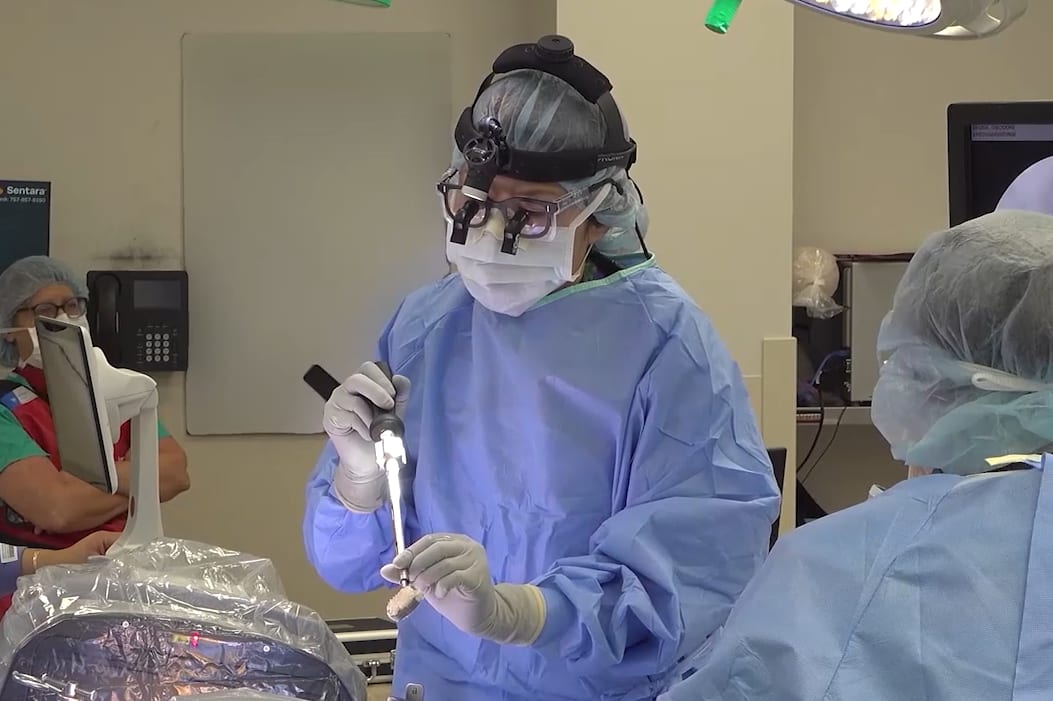
In this procedural case review, vascular surgeon Dr. Samuel Steerman and neurosurgeon Dr. Shannon Clark collaborate to perform an anterior lumbar interbody fusion (ALIF).