Redefining diagnostic lesional status in temporal lobe epilepsy with artificial intelligence
By
Ezequiel Gleichgerrcht
Erik Kaestner
Reihaneh Hassanzadeh
Rebecca W Roth
Alexandra Parashos
Kathryn A Davis
Anto Bagić
Simon S Keller
Theodor Rüber
Travis Stoub
Heath R Pardoe
Patricia Dugan
Daniel L Drane
Anees Abrol
Vince Calhoun
Ruben I Kuzniecky
Carrie R McDonald
Leonardo Bonilha
January 22, 2025
Clinical Scorecard: Utilizing Artificial Intelligence to Reassess Lesional Classification in Temporal Lobe Epilepsy Diagnosis
At a Glance
Category Detail
Condition Temporal Lobe Epilepsy (TLE) Key Mechanisms AI-based analysis of MRI detects subtle limbic and temporal atrophy patterns characteristic of TLE, including in MRI-negative patients Target Population Patients with temporal lobe epilepsy, including those classified as MRI-negative Care Setting Neurology and epilepsy diagnostic imaging centers, surgical evaluation settings
Key Highlights
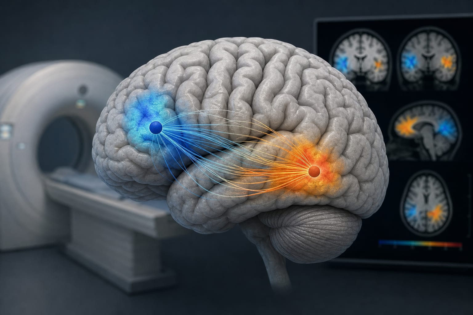
30%–50% of TLE patients are MRI-negative by human visual assessment, causing diagnostic delays. A 3D convolutional neural network (CNN) distinguished TLE from controls with 85.9% accuracy, outperforming traditional volumetric methods. AI identified MRI-negative TLE patients with 82.7% accuracy, suggesting a continuum of lesional patterns undetectable by humans. Guideline-Based Recommendations
Diagnosis
Incorporate AI-assisted MRI interpretation to improve detection of subtle lesional patterns in TLE, especially in MRI-negative cases. Use AI-derived saliency maps focusing on limbic structures (medial temporal, cingulate, orbitofrontal areas) to support diagnosis. Management
Consider AI-aided imaging findings to inform surgical candidacy and treatment planning in TLE patients. Recognize that AI can redefine 'lesional' status, potentially impacting clinical decision-making. Monitoring & Follow-up
Utilize AI tools to monitor structural brain changes over time to assess disease progression or treatment response. Risks
Be aware of potential over-reliance on AI outputs without clinical correlation. Ensure AI models are validated across diverse populations and imaging protocols to avoid misclassification. Patient & Prescribing Data
Patients with temporal lobe epilepsy, including those with MRI-negative findings
AI-enhanced imaging may reduce diagnostic uncertainty and delays, facilitating timely surgical intervention and personalized management.
Clinical Best Practices
Combine AI-assisted MRI analysis with clinical and surgical outcome data for robust TLE diagnosis. Use multicenter, large datasets to train and validate AI models for generalizability. Interpret AI saliency maps to understand neuroanatomical correlates of TLE and support clinical decisions. Maintain multidisciplinary collaboration between neurologists, radiologists, and data scientists for AI integration. References