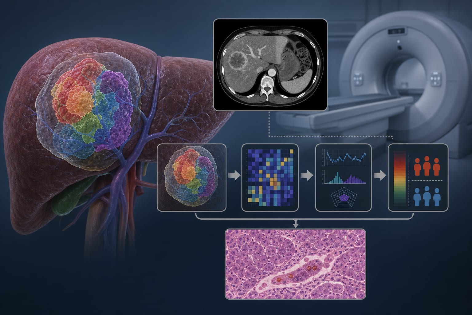
Development and validation of a radiopathomics model for predicting liver metastases of colorectal cancer
-
By
-
Han-Hui Jing
-
Di Hao
-
Xue-Jun Liu
-
Ming-Juan Cui
-
Kui-Jin Xue
-
Dong-Sheng Wang
-
Jun-Hao Zhang
-
Yun Lu
-
Guang-Ye Tian
-
Shang-Long Liu
-
December 2, 2024
-
Clinical Scorecard: Creation and assessment of a radiopathomics model for forecasting liver metastases in colorectal cancer patients
At a Glance
| Category | Detail |
|---|
| Condition | Colorectal cancer with postoperative liver metastases |
| Key Mechanisms | Integration of CT radiomics features and clinical data to predict liver metastasis risk |
| Target Population | Patients with histopathologically confirmed colorectal cancer undergoing preoperative CT |
| Care Setting | Preoperative evaluation and postoperative risk stratification in hospital or oncology centers |
Key Highlights
- Liver metastases occur in at least 50% of colorectal cancer patients postoperatively, significantly impacting mortality.
- Traditional TNM staging lacks sufficient accuracy for predicting liver metastases; radiomics offers quantitative imaging biomarkers.
- A combined model using CT radiomics and clinical features improves prediction of postoperative liver metastases.
Guideline-Based Recommendations
Diagnosis
- Use pathological examination as the gold standard for liver metastases diagnosis post-surgery.
- Employ preoperative abdominal and pelvic CT scans with standardized imaging protocols for radiomics feature extraction.
- Apply quantitative radiomics analysis following International Biomarker Standardization Initiative guidelines.
Management
- Consider preoperative treatment strategies to reduce metastasis risk based on accurate staging and risk prediction.
- Use combined radiomics and clinical models to guide personalized treatment planning and patient selection.
Monitoring & Follow-up
- Regular follow-up imaging and clinical data collection to monitor for liver metastases development postoperatively.
Risks
- Invasiveness and impracticality of frequent pathological examinations limit their use for ongoing monitoring.
- Variability in clinician experience affects the reliability of conventional CT interpretation without radiomics.
Patient & Prescribing Data
Colorectal cancer patients undergoing radical surgery with available preoperative CT and clinical data
Radiopathomics models enable early identification of high-risk patients who may benefit from intensified preoperative or adjuvant therapies.
Clinical Best Practices
- Ensure high-quality, standardized CT imaging acquisition and preprocessing for reproducible radiomics feature extraction.
- Use multidisciplinary review of imaging and clinical data to resolve discrepancies in tumor delineation.
- Apply rigorous feature selection combining statistical tests and L1 regularization to optimize predictive model performance.
- Integrate radiomics scores with clinical variables using score-level fusion methods to enhance prediction accuracy.
- Adopt models validated on retrospective cohorts with comprehensive clinical and imaging datasets.
References