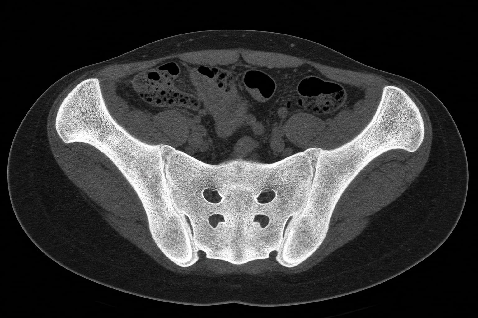
Renal osteodystrophy with near-complete bilateral sacroiliac ankylosis: a bat-like pelvic configuration on CT
-
By
-
Ramazan Orkun Onder
-
Serdar Aslan
-
July 3, 2026
-
Clinical Scorecard: Renal Osteodystrophy Accompanied by Nearly Complete Bilateral Sacroiliac Fusion: A Bat-Shaped Pelvic Appearance on CT Imaging
At a Glance
| Category | Detail |
|---|
| Condition | Renal Osteodystrophy |
| Key Mechanisms | Secondary hyperparathyroidism leading to osteosclerosis and sacroiliac abnormalities. |
| Target Population | Patients with chronic kidney disease stage 5D on hemodialysis. |
| Care Setting | Clinical evaluation and imaging in nephrology. |
Key Highlights
- Patient presented with abdominal pain radiating to the lower back.
- CT imaging revealed bilateral renal atrophy and nephrolithiasis.
- Near-complete bilateral sacroiliac ankylosis observed on pelvic CT.
- Biochemical evaluation indicated severe secondary hyperparathyroidism.
- Negative HLA-B27 and absence of spondyloarthritis features.
Guideline-Based Recommendations
Diagnosis
- Consider renal osteodystrophy in patients with chronic kidney disease presenting with skeletal abnormalities.
Management
- Monitor biochemical markers such as parathyroid hormone, calcium, and phosphate in patients with chronic kidney disease.
Monitoring & Follow-up
- Regular imaging and biochemical evaluations to assess skeletal involvement and mineral metabolism.
Risks
- Increased risk of skeletal abnormalities and complications in patients with chronic kidney disease.
Patient & Prescribing Data
Patients with chronic kidney disease stage 5D on hemodialysis.
Management of secondary hyperparathyroidism is crucial in preventing skeletal complications.
Clinical Best Practices
- Utilize CT imaging to assess skeletal manifestations in renal osteodystrophy.
- Evaluate for secondary hyperparathyroidism in patients with chronic kidney disease.
Related Resources & Content