Super-resolution for localizing electrode grids as small, deformable objects during epilepsy surgery using augmented reality headsets - Scorecard - MDSpire
Advertisement
Super-resolution for localizing electrode grids as small, deformable objects during epilepsy surgery using augmented reality headsets
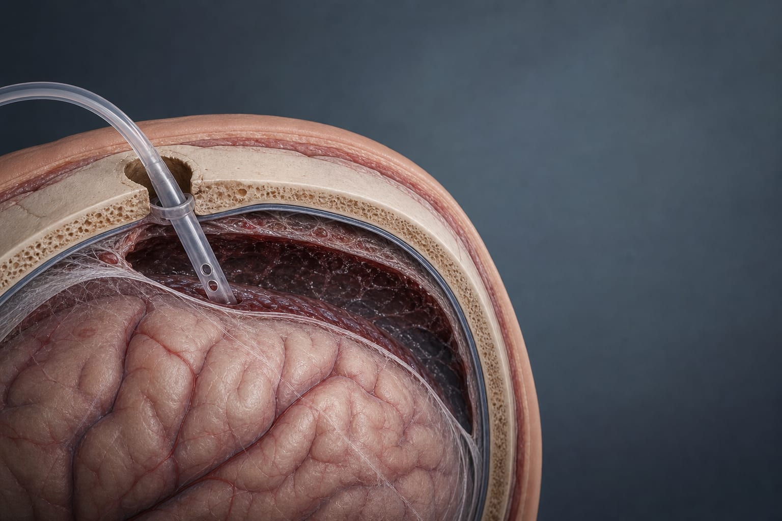
Intraoperative electrocorticography (ioECoG) grid placement and localization to delineate epileptic tissue
Target Population
Patients with focal epilepsy refractory to anti-seizure medication
Care Setting
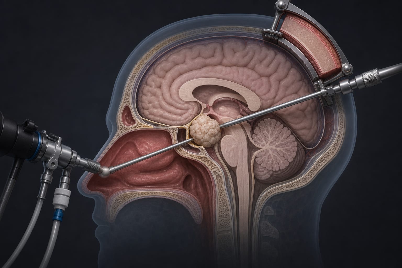
Neurosurgical operating room during tailored epilepsy surgery
Key Highlights
Precise localization of deformable ioECoG grids is critical for successful epilepsy surgery outcomes.
Current localization methods rely on visual inspection or photograph-based techniques with limitations in accuracy, time, and expertise required.
Augmented reality head-mounted displays combined with AI techniques offer a novel approach for real-time, accurate, and efficient ioECoG grid localization.
Guideline-Based Recommendations
Diagnosis
Use intraoperative electrocorticography (ioECoG) grids placed on the cortex to localize epileptic tissue.
Employ presurgical 3D MRI for anatomical reference in grid localization.
Management
Consider epilepsy surgery for patients with focal refractory epilepsy unresponsive to medication.
Utilize augmented reality (AR) head-mounted displays integrated with AI for enhanced localization of ioECoG grids during surgery.
Monitoring & Follow-up
Monitor ioECoG grid placement accuracy intraoperatively to ensure complete resection of epileptic tissue.
Compare localization results against established tracking systems (e.g., commercial NDI tracking) as ground truth.
Risks
Inaccurate localization of ioECoG grids may lead to incomplete resection and suboptimal surgical outcomes.
Prolonged registration times and need for specialized expertise may delay surgery and increase risk.
Patient & Prescribing Data
Patients with focal epilepsy refractory to anti-seizure medications undergoing epilepsy surgery
Accurate localization of epileptic tissue via ioECoG grids improves surgical success; AR and AI integration may enhance precision and workflow.
Clinical Best Practices
Integrate AR HMDs with AI-based object detection, super-resolution, and 2D pose estimation to improve ioECoG grid localization accuracy.
Correct camera image distortion and apply image rectification prior to grid detection to enhance triangulation accuracy.
Use synthetic data to train AI models to overcome medical data scarcity challenges.
Aim for localization accuracy within clinically acceptable thresholds (mean error <5 mm).
Prefer hands-free AR HMDs over bulky or workflow-disruptive devices for intraoperative use.