Comparative analysis of the lumboperitoneal shunt versus ventriculoperitoneal shunt for leptomeningeal metastasis-associated hydrocephalus in non-small cell lung cancer - Scorecard - MDSpire
Advertisement
Comparative analysis of the lumboperitoneal shunt versus ventriculoperitoneal shunt for leptomeningeal metastasis-associated hydrocephalus in non-small cell lung cancer
Clinical Scorecard: Evaluation of Lumboperitoneal Shunt Versus Ventriculoperitoneal Shunt in Managing Hydrocephalus Due to Leptomeningeal Metastasis in Patients with Non-Small Cell Lung Cancer
At a Glance
Category
Detail
Condition
Hydrocephalus caused by leptomeningeal metastasis in NSCLC patients
Key Mechanisms
CSF flow obstruction by malignant cells causing increased intracranial pressure
Target Population
Adult patients (≥18 years) with NSCLC and confirmed leptomeningeal metastasis-associated communicating hydrocephalus
Care Setting
Neurosurgical and oncology clinical settings with capability for shunt surgery
Key Highlights
Leptomeningeal metastasis is a rare, aggressive complication of advanced NSCLC with poor prognosis.
LM-associated hydrocephalus occurs in 1%–5% of LM patients, causing neurological symptoms due to raised ICP.
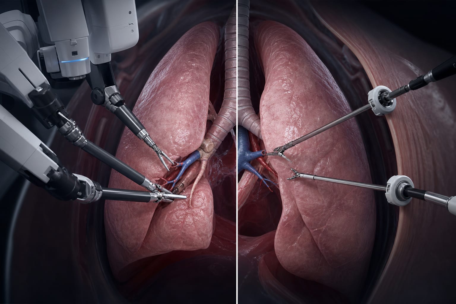
LP shunt surgery is a less invasive alternative to VP shunt with potential benefits including lower complication risk and avoidance of general anesthesia.
Guideline-Based Recommendations
Diagnosis
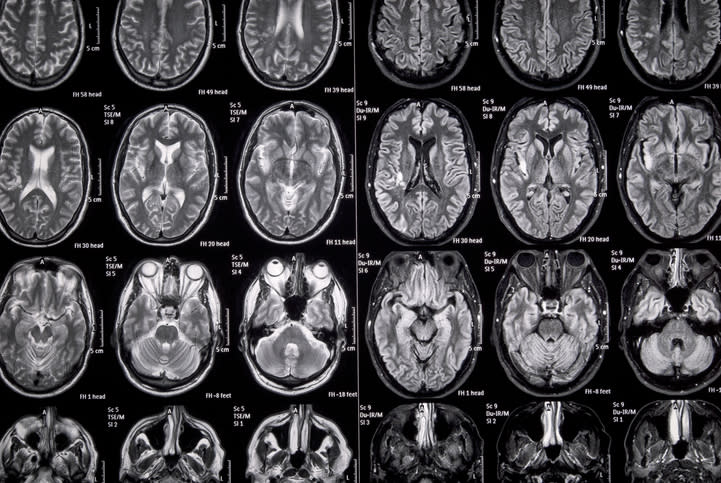
Confirm LM diagnosis using gadolinium-enhanced brain MRI, CSF cytology or meningeal biopsy, and clinical signs per EANO-ESMO guidelines.
Classify LM based on MRI enhancement patterns and cytology results (types 1A to 2D).
Consider VP shunt or endoscopic third ventriculostomy to relieve increased ICP in LM-associated hydrocephalus.
LP shunt surgery is a viable alternative for communicating hydrocephalus, especially in patients unsuitable for general anesthesia.
Palliative care or lumbar drainage may be options when surgery is contraindicated.
Monitoring & Follow-up
Monitor neurological symptoms and imaging findings post-shunt surgery to assess ICP control and shunt function.
Regular follow-up imaging and clinical evaluation to detect shunt complications or hydrocephalus progression.
Risks
VP shunt surgery involves risks related to ventricular catheter insertion through brain tissue.
LP shunt may reduce surgical risks but requires careful patient selection to exclude obstructive hydrocephalus.
Poor patient condition may limit surgical options and affect prognosis.
Patient & Prescribing Data
Adults with NSCLC and confirmed LM-associated communicating hydrocephalus eligible for surgical intervention
LP shunts may offer safer, less invasive management with comparable effectiveness to VP shunts; however, evidence is limited and further comparative studies are needed.
Clinical Best Practices
Use multidisciplinary evaluation including oncology and neurosurgery specialists to determine surgical eligibility.
Follow EANO-ESMO guidelines for LM diagnosis and classification to guide treatment decisions.
Select shunt type based on hydrocephalus subtype, patient condition, and surgical risk profile.
Ensure thorough preoperative assessment and postoperative monitoring to optimize outcomes.
by Minjoon Kim, Chaejin Lee, Sang-Youl Yoon, Seong-Hyun Park, Jeong-Hyun Hwang, Kyunghun Kang, Eunhee Park, Sunha Choi, Shin Yup Lee, Seung Soo Yoo, Yee Soo Chae, Ki-Su Park