Sinonasal and skull base phosphaturic mesenchymal tumours: a case series and narrative review
-
By
-
Genwang Pei
-
Rongfeng Lin
-
Guangqi Li
-
Yinyan Lai
-
June 18, 2026
-
Clinical Scorecard: Phosphaturic Mesenchymal Tumours in the Sinonasal Region and Skull Base: A Case Series with Comprehensive Review
At a Glance
| Category | Detail |
|---|
| Condition | Phosphaturic Mesenchymal Tumours (PMTs) |
| Key Mechanisms | Tumour cell secretion of fibroblast growth factor 23 (FGF23) inhibits phosphate reabsorption, leading to hypophosphataemia and osteomalacia. |
| Target Population | Adults aged 28 to 67 years with sinonasal or skull base tumours. |
| Care Setting | Retrospective analysis in a clinical institution. |
Key Highlights
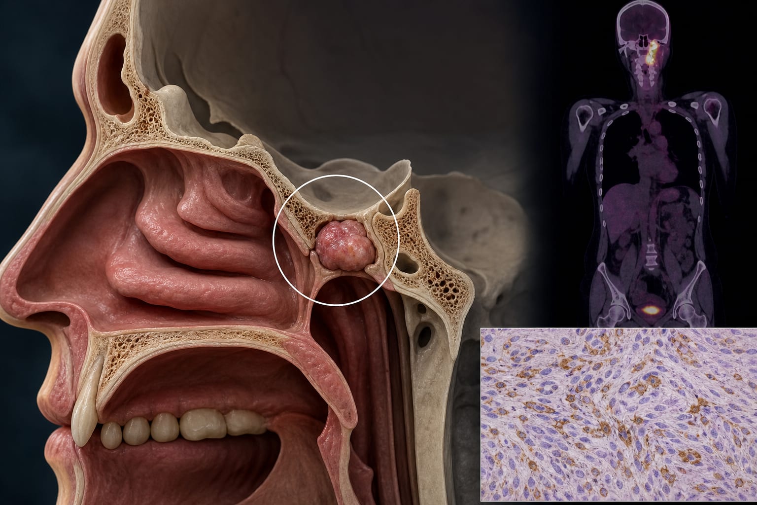
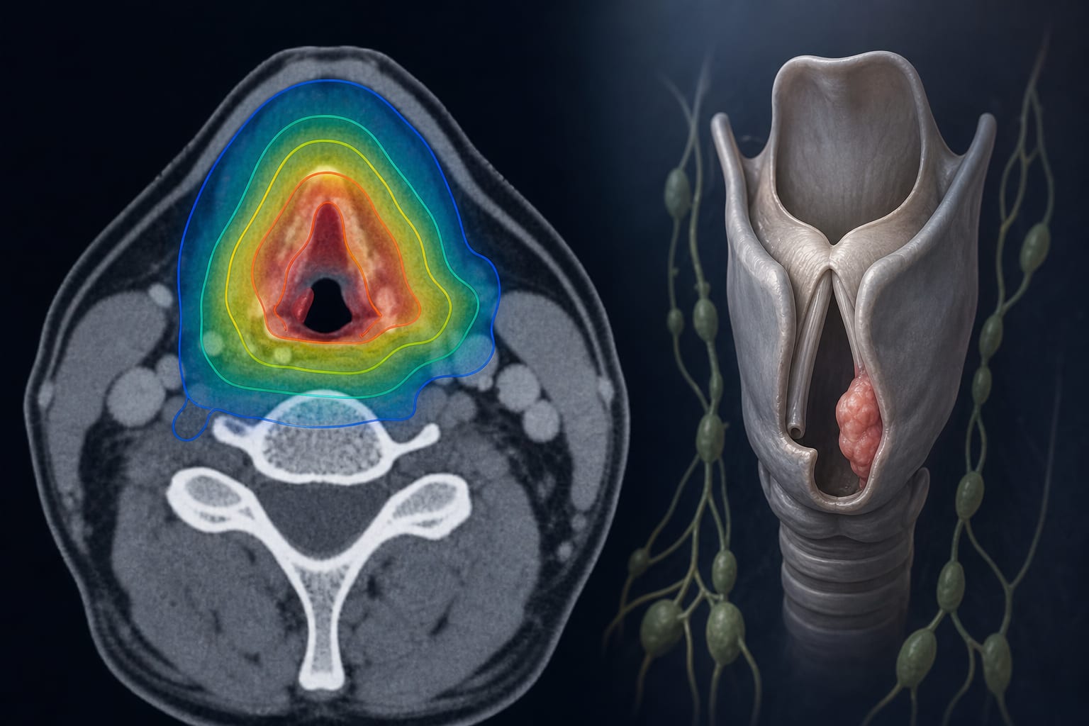
- PMTs are rare in the sinonasal region and skull base, often leading to diagnostic challenges.
- Persistent hypophosphataemia is a vital diagnostic indicator.
- PET imaging showing somatostatin receptor positivity is a key diagnostic tool.
- All patients underwent surgical resection, with postoperative serum phosphorus levels normalizing within 4–10 days.
- Four out of five patients experienced diagnostic delays, with misdiagnoses including nasal polyps.
Guideline-Based Recommendations
Diagnosis
- Utilize PET imaging for diagnosis, particularly for somatostatin receptor expression.
- Monitor serum phosphorus levels for diagnostic confirmation.
Management
- Surgical resection is the primary treatment for PMTs.
Monitoring & Follow-up
- Postoperative monitoring of serum phosphorus levels to evaluate cure and recurrence.
Risks
- High rates of missed and misdiagnosis due to atypical clinical manifestations and tumour obscurity.
Patient & Prescribing Data
Five patients aged 28 to 67 years with confirmed sinonasal/skull base PMTs.
All patients underwent endoscopic surgery; one patient experienced recurrence after 2 years.
Clinical Best Practices
- Consider PMTs in differential diagnoses for patients with unexplained hypophosphataemia and bone pain.
- Employ comprehensive imaging techniques, including CT, MRI, and PET, for accurate diagnosis.
Related Resources & Content