Single-step 3D printing aided cranio-orbital reconstruction with patient specific polyetheretherketone implants after resection of benign spheno-orbital tumors - Scorecard - MDSpire
Clinical Scorecard: Three-Dimensional Printing Facilitated Cranio-Orbital Reconstruction Using Customized Polyetheretherketone Implants Following Benign Spheno-Orbital Tumor Resection
At a Glance
Category
Detail
Condition
Benign spheno-orbital tumors with anterior skull base infiltration
Key Mechanisms
Complex tumor resection and rigid cranio-orbital reconstruction using patient-specific CAD/CAM polyetheretherketone (PEEK) implants facilitated by 3D printing and virtual surgical planning
Target Population
Patients with benign spheno-orbital tumors requiring single-stage tumor resection and cranio-orbital reconstruction
Care Setting
Academic teaching hospital with interdisciplinary skull base team (neurosurgery and maxillofacial surgery)
Key Highlights
Rigid orbital reconstruction with customized PEEK implants prevents malalignment and pulsating eye bulb, preserving orbital volume and reducing exophthalmos.
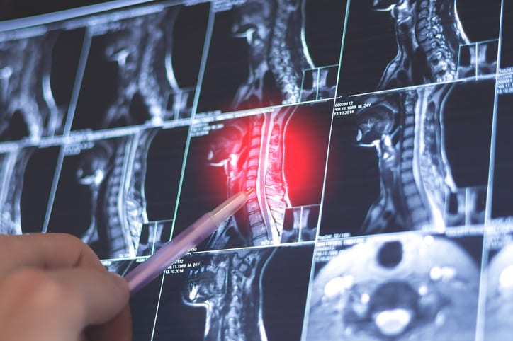
Preoperative virtual planning integrates CT, MRI, and PET imaging to define tumor and bone resection margins, enabling precise design of cutting guides and implants.
Single-step surgical approach combining tumor resection and immediate cranio-orbital reconstruction using 3D printed patient-specific implants improves functional, visual, and aesthetic outcomes.
Guideline-Based Recommendations
Diagnosis
Use thin-sliced 1 mm cranial CT, contrast-enhanced brain MRI, and optionally 68Ga-DOTATOC PET/CT for detailed tumor and bone margin delineation.
Define tumor and safe skull resection margins collaboratively by neurosurgeons and maxillofacial surgeons using advanced planning software.
Management
Perform single-stage microsurgical tumor resection and cranio-orbital reconstruction under general anesthesia.
Utilize CAD/CAM designed PEEK patient-specific implants and cutting guides produced by 3D printing for precise reconstruction.
Intraoperatively adapt implants as needed to accommodate actual resection margins.
Verify extent of resection and implant placement with intraoperative imaging.
Monitoring & Follow-up
Conduct postoperative cranial MRI or CT imaging on day one and at 3 to 6 months post-surgery.
Perform regular ophthalmological examinations to assess visual function and monitor for exophthalmos.
Calculate Exophthalmic Index preoperatively and postoperatively to evaluate orbital volume restoration.
Risks
Potential deterioration of visual acuity due to optic nerve traction during surgery.
Challenges in preoperative planning due to complex tumor margins and bone cuts.
Possible need for intraoperative implant modification if resection margins differ from planned.
Patient & Prescribing Data
Eleven consecutive female patients aged 19–72 years with benign spheno-orbital tumors undergoing single-stage resection and reconstruction.
Majority presented with exophthalmos; some had prior resections, biopsies, or radiotherapy. Median surgery time was approximately 13 hours. Interdisciplinary surgical collaboration and advanced imaging guided personalized implant design and successful reconstruction.
Clinical Best Practices
Engage an interdisciplinary skull base team including neurosurgeons and maxillofacial surgeons for optimal tumor control and reconstruction.
Employ advanced imaging modalities and digital planning software for precise definition of tumor and bone resection margins.
Use 3D printing at point of care for surgical planning models and patient-specific cutting guides and implants.
Perform intraoperative imaging to confirm resection extent and implant positioning.
Obtain informed consent and adhere to ethical standards including institutional and Helsinki declarations.
by Arwin Rezai, Johannes P. Pöppe, Alexander Gaggl, Christoph J. Griessenauer, Christoph Schwartz, Herbert Krainz, Moritz Ueberschaer, Petra A. Mercea, Simon Enzinger
For decades, treating pituitary tumors often meant a craniotomy. Large incisions, brain retraction, and longer recovery times were part of the equation, even for benign tumors. That is no longer the standard.
A prospective cohort study suggests that clinically meaningful quality‐of‐life changes continue well beyond the first postoperative year, with one‐year complications emerging as the strongest potentially modifiable predictor of later decline.