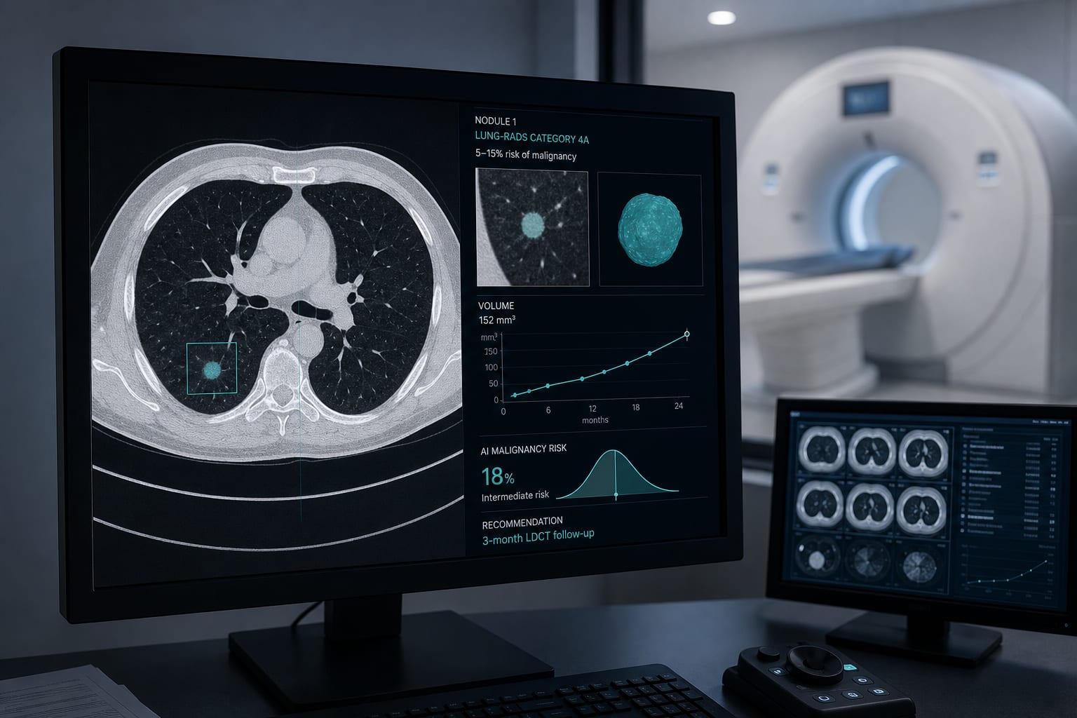
Clinical Scorecard: Analysis of 3D Fractal Dimensions in CT Imaging for Predicting Microvascular Invasion in Hepatocellular Carcinoma
At a Glance
Category
Detail
Condition
Hepatocellular carcinoma (HCC) with microvascular invasion (MVI)
Key Mechanisms
Microvascular invasion involves tumor cell presence in small vessels, influencing tumor recurrence and prognosis; fractal analysis quantifies tumor morphological complexity related to vascularization
Preoperative imaging assessment and surgical planning in tertiary medical centers
Key Highlights
MVI is a critical histopathological factor linked to poor prognosis and tumor recurrence in HCC.
Preoperative identification of MVI is limited by reliance on surgical specimens; current radiological and clinical markers have subjective limitations.
Fractal dimension analysis of 3D tumor morphology on contrast-enhanced CT offers an objective quantitative method to assess microvascular complexity and predict MVI status.
Guideline-Based Recommendations
Diagnosis
Definitive MVI diagnosis requires histopathological examination of surgical specimens.
Preoperative prediction of MVI can be enhanced by assessing tumor morphology using fractal dimension analysis on contrast-enhanced CT.
Consider integrating fractal analysis with clinical factors (tumor size, number, AFP levels) for improved MVI risk estimation.
Management
Patients with predicted MVI may require wider surgical resection margins.
Neoadjuvant or adjuvant therapies should be considered in HCC patients with MVI to reduce recurrence risk.
MVI status influences eligibility and prognosis in liver transplantation decisions.
Monitoring & Follow-up
Postoperative survival outcomes should be monitored in relation to preoperative fractal dimension findings.
Regular imaging follow-up is recommended to detect tumor recurrence, especially in patients with MVI.
Risks
Subjective interpretation of radiological features can lead to interobserver variability.
Exclusion of patients with prior HCC treatments or macroscopic vascular invasion is necessary to avoid confounding fractal analysis results.
Patient & Prescribing Data
HCC patients undergoing curative resection with available preoperative contrast-enhanced CT imaging
Fractal dimension analysis may guide surgical planning and adjuvant therapy decisions by predicting MVI presence, potentially improving individualized treatment strategies.
Clinical Best Practices
Standardize CT imaging protocols and preprocessing (resampling, intensity normalization) for consistent fractal analysis.
Use 3D tumor segmentation on portal venous phase images focusing on the largest tumor in multifocal cases.
Employ fractal dimension box-counting methods with curve-fitting techniques to quantify tumor morphological complexity.
Combine fractal analysis results with clinical and pathological data for comprehensive MVI risk assessment.
Ensure reproducibility by validating inter-reader and intra-reader segmentation consistency.