The Role of Non-Contrast CT Attenuation Values in Predicting Hematoma Expansion in Spontaneous Intracerebral Hemorrhage
By
Jing Wang
Chuyue Wu
Jing Guo
Zhenjie Yang
Lei He
Shengli Chen
Lina Zhang
July 17, 2026
Clinical Scorecard: The Role of Non-Contrast CT Attenuation Values in Predicting Hematoma Expansion in Spontaneous Intracerebral Hemorrhage
At a Glance
Category Detail
Condition Spontaneous Intracerebral Hemorrhage (sICH) Key Mechanisms Relative CT attenuation value predicts hematoma expansion risk. Target Population Patients aged 18 years or older with sICH treated with minimally invasive surgery. Care Setting Department of Neurology at Chongqing University Three Gorges Hospital
Key Highlights
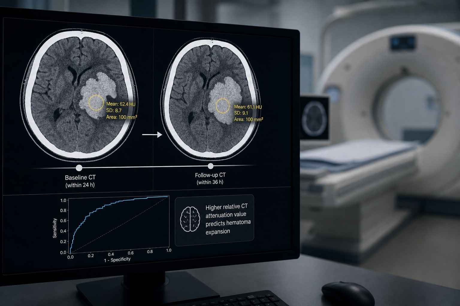
25.9% of patients with sICH exhibited hematoma expansion (HE). Relative CT attenuation value of 33.35 Hounsfield units (HUs) is a critical threshold for predicting HE. The area under the ROC curve (AUC) for predicting HE was 0.665. Sensitivity and specificity for a relative CT attenuation value of ≤33.35 HU were 40.9% and 83.1%, respectively. Early identification of HE risk could facilitate timely interventions. Guideline-Based Recommendations
Diagnosis
Baseline CT scan within 24 hours of symptom onset. Follow-up CT scan within 36 hours. Management
Consider relative CT attenuation values in treatment planning. Monitoring & Follow-up
Monitor for hematoma expansion within the first 24 hours. Risks
High mortality and morbidity rates associated with sICH. Patient & Prescribing Data
Patients with spontaneous intracerebral hemorrhage treated at Chongqing University Three Gorges Hospital.
Minimally invasive surgery is a treatment option for eligible patients.
Clinical Best Practices
Utilize relative CT attenuation values to assess risk of hematoma expansion. Implement early imaging protocols for timely diagnosis and management. Related Resources & Content