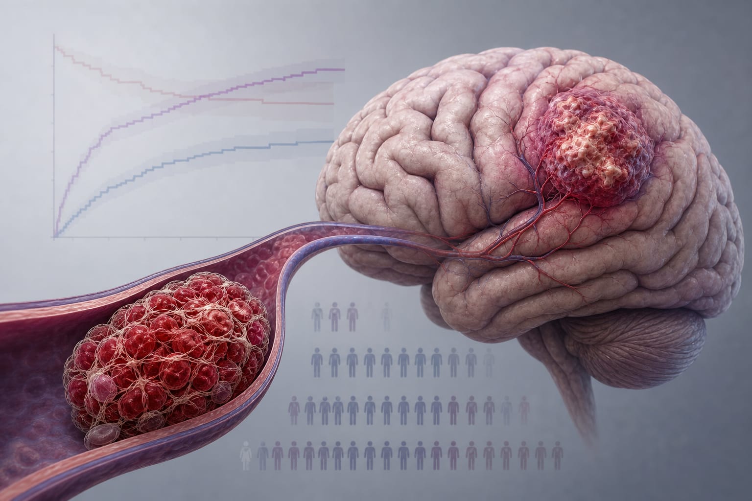
Neurosurgical and neuro-oncology clinical settings with preoperative MRI imaging and postoperative functional assessment
Key Highlights
High-grade gliomas represent 60–70% of new brain tumor cases with median survival around 14 months despite standard treatment.
RS-fMRI enables functional brain network mapping without task performance, suitable even under sedation, with lower failure rates than task-based fMRI.
Machine learning models using RS-fMRI connectivity, tumor overlap with networks, tumor volume, and age can accurately predict postoperative functional outcomes (KPS ≥ 70 vs < 70).
Guideline-Based Recommendations
Diagnosis
Use histopathological and immunohistochemical criteria per WHO guidelines for definitive HGG diagnosis.
Acquire pre-surgical structural MRI (T1, T2, DTI) and resting state fMRI to evaluate tumor characteristics and brain functional networks.
Management
Perform maximal safe gross total resection followed by radiation and adjuvant chemoradiotherapy.
Incorporate RS-fMRI data into surgical planning to balance maximal tumor resection with functional preservation.
Monitoring & Follow-up
Assess postoperative functional status using Karnofsky Performance Scale (KPS) to evaluate patient outcomes.
Surgical resection may cause or worsen functional impairments impacting quality of life and overall survival.
Limitations of task-based fMRI include patient inability to perform tasks; RS-fMRI offers an alternative with fewer constraints.
Patient & Prescribing Data
Adults with newly diagnosed high-grade gliomas undergoing biopsy or surgical resection with available preoperative MRI data.
Machine learning models integrating RS-fMRI and clinical features can inform prognosis and guide surgical decision-making to optimize functional outcomes.
Clinical Best Practices
Utilize resting state fMRI for functional brain mapping preoperatively, especially in patients unable to perform task-based fMRI.
Apply machine learning techniques to extract relevant imaging and clinical features for individualized outcome prediction.
Balance extent of tumor resection with preservation of critical functional networks to improve postoperative quality of life.
by Patrick H. Luckett, Michael O. Olufawo, Ki Yun Park, Bidhan Lamichhane, Donna Dierker, Gabriel Trevino Verastegui, John J. Lee, Peter Yang, Albert Kim, Omar H. Butt, Milan G. Chheda, Abraham Z. Snyder, Joshua S. Shimony, Eric C. Leuthardt