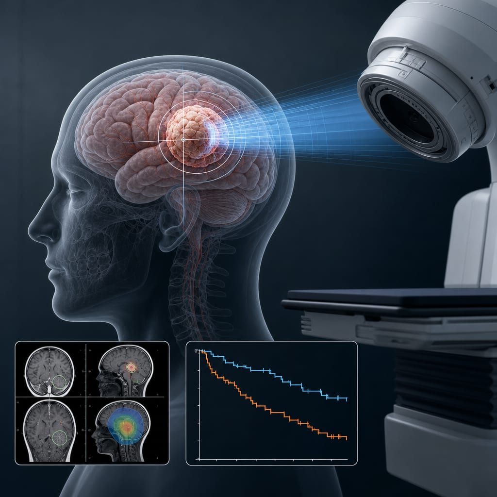
Intra-cavitary radiotherapy for surgically resected brain metastases: a comprehensive analysis including an individual patient data meta-analysis of intraoperative radiotherapy (IORT) and brachytherapy (IBT) - Scorecard - MDSpire
Advertisement
Intra-cavitary radiotherapy for surgically resected brain metastases: a comprehensive analysis including an individual patient data meta-analysis of intraoperative radiotherapy (IORT) and brachytherapy (IBT)
Clinical Scorecard: Evaluation of Intra-cavitary Radiotherapy Techniques for Resected Brain Metastases: An In-depth Review with Individual Patient Data Meta-analysis of Intraoperative Radiotherapy (IORT) and Brachytherapy (IBT)
At a Glance
Category
Detail
Condition
Resected brain metastases
Key Mechanisms
Localized intra-cavitary radiation delivered via intraoperative radiotherapy (IORT) using low-energy X-rays or electrons, and intracavitary brachytherapy (IBT) using implanted radioactive sources
Target Population
Adult patients with histologically confirmed brain metastases undergoing surgical resection
Care Setting
Neurosurgical and radiation oncology settings for postoperative localized radiotherapy
Key Highlights
Brain metastases affect approximately 2% of all cancer patients and up to 12.1% of those with metastatic disease at diagnosis.
Postoperative localized radiotherapy with IORT or IBT reduces treatment delays and limits radiation exposure to healthy brain tissue, achieving 1-year local control rates of 85–96%.
IORT and IBT show promising efficacy and safety profiles in improving local control and preserving cognitive function compared to whole-brain radiotherapy.
Guideline-Based Recommendations
Diagnosis
Histological confirmation of brain metastases prior to surgical resection.
Use of imaging and clinical assessment to identify accessible lesions suitable for maximal safe resection.
Management
Maximal safe surgical resection for symptomatic or accessible brain metastases.
Adjuvant localized radiotherapy via IORT or IBT immediately during or after resection to reduce microscopic residual disease and recurrence.
Avoidance of whole-brain radiotherapy when possible to minimize neurocognitive decline.
Monitoring & Follow-up
Regular follow-up imaging to assess local control and detect distant brain failure.
Monitoring for treatment-related toxicities including radiation necrosis and leptomeningeal disease.
Assessment of wound healing and complications post-radiotherapy.
Risks
Potential for radiation necrosis and leptomeningeal disease, though rates are reported and monitored.
Risk of wound complications associated with intra-cavitary radiotherapy procedures.
Selection bias and heterogeneity in study populations may affect outcome interpretation.
Patient & Prescribing Data
858 adult patients with resected brain metastases treated with IORT or IBT across 23 studies (1999–2024).
Both IORT and IBT demonstrate high 1-year local control rates (85–96%) with reduced treatment delays and preservation of cognitive function; patient selection and treatment modality should consider tumor characteristics and prior therapies.
Clinical Best Practices
Employ maximal safe resection for accessible brain metastases to enable cytoreduction and histological diagnosis.
Implement intraoperative or immediate postoperative localized radiotherapy (IORT or IBT) to target residual microscopic disease and improve local control.
Prefer localized radiotherapy techniques over whole-brain radiotherapy to minimize neurocognitive side effects.
Use standardized protocols for patient selection, treatment delivery, and follow-up to optimize outcomes and monitor for toxicity.
Incorporate multidisciplinary evaluation including neurosurgery, radiation oncology, and neuro-oncology for individualized treatment planning.