High-frequency ultrasound features of cutaneous hidrocystoma: a case series with imaging-pathology correlation
By
Lina Kong
Xiao Tang
Kunming Pu
Xiachuan Qin
June 26, 2026
Clinical Scorecard: Ultrasound Characteristics of Cutaneous Hidrocystoma: A Case Series with Imaging and Pathology Correlation
At a Glance
Category Detail
Condition Cutaneous Hidrocystoma Key Mechanisms Benign cystic lesion of sweat gland origin Target Population Middle-aged and older adults Care Setting Dermatology practice
Key Highlights
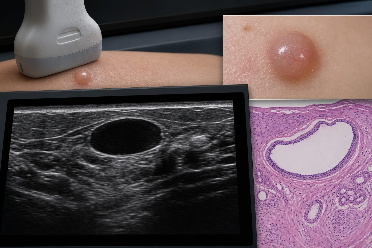
High-frequency ultrasound (HFUS) provides additional information for lesion characterization. Eleven patients with pathologically confirmed hidrocystoma were analyzed. Most lesions were located in the head and neck region and measured less than 1.5 cm. Sonographic appearances included heterogeneous echogenicity and anechoic cystic features. HFUS aids in preoperative localization and narrowing differential diagnosis. Guideline-Based Recommendations
Diagnosis
Clinical examination by dermatologists is the first-line diagnostic approach. Dermoscopy is increasingly used as a non-invasive adjunctive tool. Management
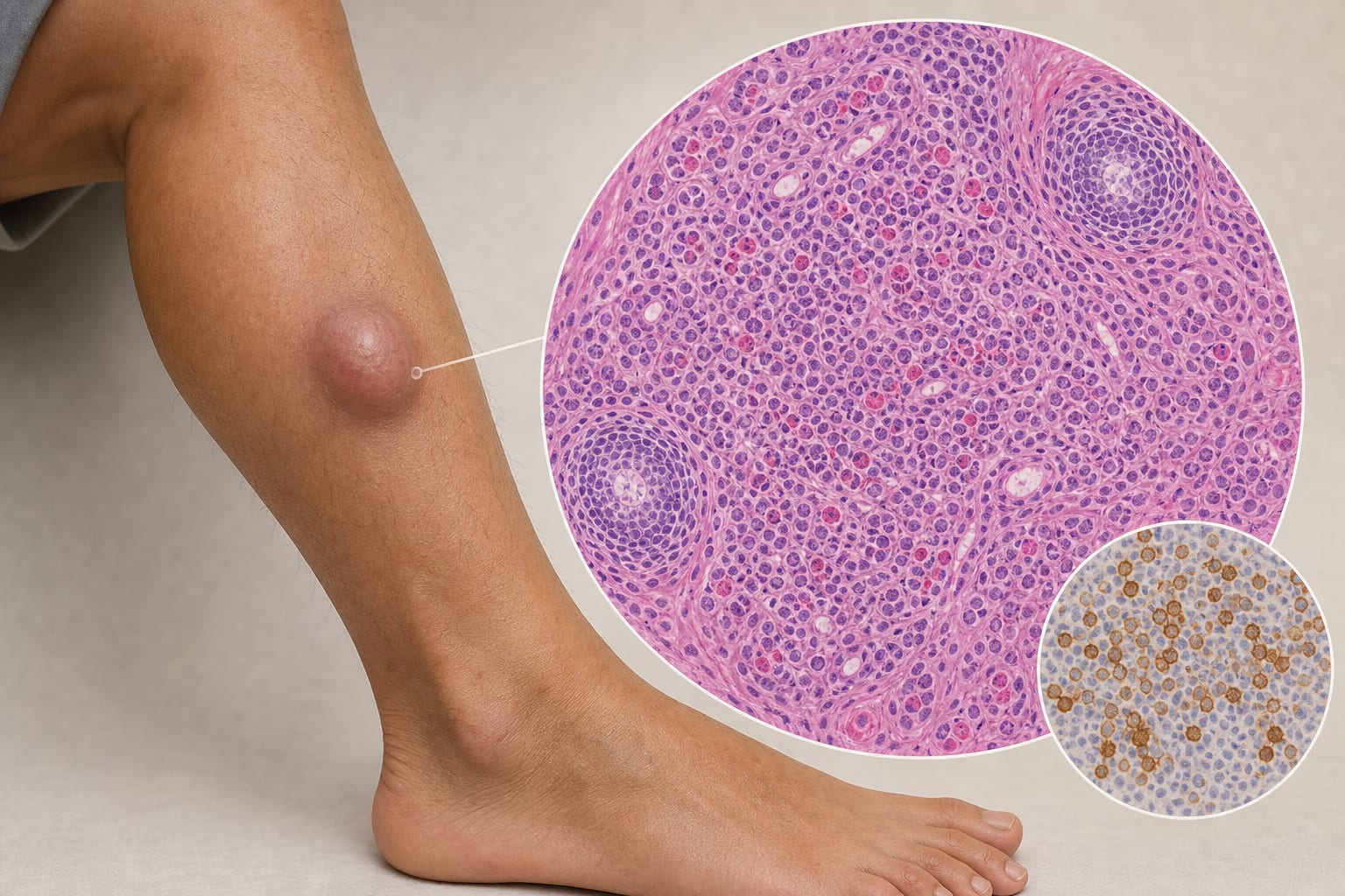
Histopathologic examination remains the reference standard for definitive diagnosis. Monitoring & Follow-up
Preoperative ultrasound findings should be correlated with postoperative histopathologic results. Risks
Invasive nature of histopathologic examination limits its routine preoperative use. Patient & Prescribing Data
Patients with pathologically confirmed cutaneous hidrocystoma.
HFUS allows precise delineation of lesion depth and vascular relationships.
Clinical Best Practices
Utilize HFUS for detailed evaluation of superficial soft-tissue lesions. Consider dermoscopy as a complementary tool for assessing surface features. Related Resources & Content