Differential infiltration of CD4+ and CD8+ T cells and expression of PD-L1 in paired biopsy and resection specimens of gastric and colorectal adenocarcinomas - Scorecard - MDSpire
Advertisement
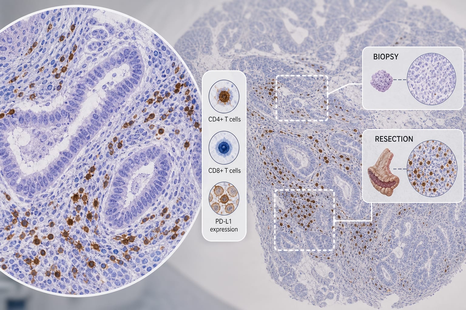
Differential infiltration of CD4+ and CD8+ T cells and expression of PD-L1 in paired biopsy and resection specimens of gastric and colorectal adenocarcinomas
Clinical Scorecard: Comparative Analysis of CD4+ and CD8+ T Cell Infiltration and PD-L1 Expression in Biopsy Versus Resection Samples of Gastric and Colorectal Adenocarcinomas
At a Glance
Category
Detail
Condition
Gastric and Colorectal Adenocarcinomas
Key Mechanisms
CD4+ T cell and CD8+ T cell infiltration, PD-L1 expression
Target Population
Patients with gastric and colorectal adenocarcinomas
Care Setting
Oncology
Key Highlights
Higher CD4+ and CD8+ T cell density and PD-L1 expression in resection specimens compared to biopsies in gastric adenocarcinoma.
In colorectal adenocarcinoma, only CD8+ T cell density was significantly higher in resection specimens.
CD4+ T cells negatively correlated with Ki-67 in colorectal adenocarcinoma.
Preoperative neutrophil-to-lymphocyte ratio positively correlated with tumor diameter in both cancers.
Guideline-Based Recommendations
Diagnosis
Assess CD4+ and CD8+ T cell density and PD-L1 expression in resection specimens.
Management
Consider tumor-type-specific immune assessment of biopsies.
Monitoring & Follow-up
Monitor correlations between immune cell infiltration and tumor markers.
Risks
Biopsy specimens may not accurately reflect the immune microenvironment.
Patient & Prescribing Data
Patients undergoing treatment for gastric and colorectal adenocarcinomas.
Clinical Best Practices
Utilize resection specimens for more accurate immune profiling.
Evaluate immune cell ratios at tumor center and invasive margin.