Clinical Scorecard: Evaluating the Precision of Probabilistic Tractography in Identifying Optimal Targets for Deep Brain Stimulation: Addressing the Discrepancies
At a Glance
Category
Detail
Condition
Essential Tremor (ET) and Parkinson's Disease (PD) treated with Deep Brain Stimulation (DBS)
Key Mechanisms
Probabilistic tractography based on Diffusion Tensor Imaging (DTI) to delineate white matter fiber tracts and identify patient-specific DBS targets
Target Population
Patients undergoing DBS surgery for ET and PD
Care Setting
Neurosurgical centers performing DBS with advanced neuroimaging and image processing
Three main tractography approaches in DBS: distance analysis to fiber tracts, fiber visualization from electrode poles, and connectivity-based localization of targets not visible on conventional MRI.
Workflow steps including thresholding for fiber binarization, manual vs automated distance measurements, and normalization to MNI space introduce measurable errors impacting electrode-to-tract distance assessments.
Guideline-Based Recommendations
Diagnosis
Use preoperative 3T MRI with DTI sequences (64 gradient directions, 2 mm isotropic voxels) under general anesthesia to minimize motion artifacts.
Acquire antiparallel b-zero images to estimate susceptibility-induced distortions.
Management
Apply probabilistic tractography workflows (e.g., FSL and LeadDBS) for patient-specific DBS target identification.
Consider connectivity-based localization for targets not visible on conventional MRI such as the ventral intermediate nucleus (VIM).
Monitoring & Follow-up
Evaluate and quantify errors introduced by thresholding, manual measurements, and normalization steps to ensure accuracy in electrode placement relative to fiber tracts.
Risks
Potential inaccuracies in DBS electrode targeting due to variability in tractography processing steps and manual interventions.
Patient & Prescribing Data
40 patients (22 with PD implanted in STN, 18 with ET implanted in VIM)
Probabilistic tractography can inform electrode placement relative to dentato-rubro-thalamic tract components associated with tremor reduction, but requires careful error assessment in workflow steps.
Clinical Best Practices
Standardize imaging acquisition protocols including use of antiparallel b-zero images to reduce susceptibility artifacts.
Use probabilistic rather than deterministic tractography to capture complex fiber anatomy relevant for DBS targeting.
Incorporate automated distance measurement tools to reduce manual measurement errors.
Normalize imaging data to standard spaces (e.g., MNI) cautiously, acknowledging introduced spatial errors.
Perform rigorous error assessment at each workflow step to optimize clinical applicability of tractography findings.
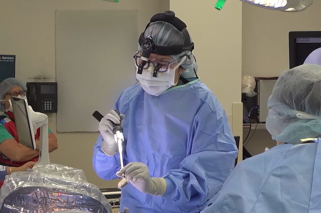
In this procedural case review, vascular surgeon Dr. Samuel Steerman and neurosurgeon Dr. Shannon Clark collaborate to perform an anterior lumbar interbody fusion (ALIF).
Epilepsy remains a life-altering condition, particularly due to the unpredictable nature of seizures and their cumulative impact on cognition, independence and quality of life.