Optimizing left atrial appendage imaging: the diagnostic value of left lateral decubitus cardiac CT angiography
-
By
-
Zihao Wang
-
ChenJing Wu
-
Yi Mang
-
Zhuang Zhuang
-
Xinshi Huang
-
Zhenzhang Wang
-
June 5, 2026
-
Clinical Scorecard: Enhancing Imaging of the Left Atrial Appendage: Assessing the Diagnostic Efficacy of Left Lateral Decubitus Cardiac CT Angiography
At a Glance
| Category | Detail |
|---|
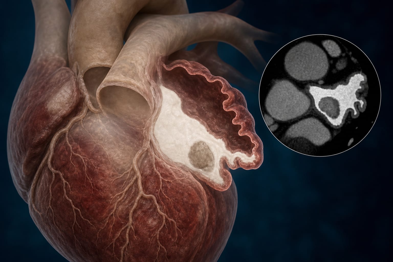
| Condition | Left Atrial Appendage Thrombus Detection |
| Key Mechanisms | Single-phase left lateral decubitus cardiac CT angiography (LLD-CCTA) improves contrast filling and reduces radiation dose. |
| Target Population | Patients with atrial fibrillation undergoing assessment for LAA thrombus. |
| Care Setting | Single-center retrospective study. |
Key Highlights
- LLD-CCTA achieved 100% sensitivity and 94% specificity for thrombus detection.
- Mean effective radiation dose reduced from 7.84 mSv to 3.89 mSv.
- Early filling defect rate decreased from 28% to 8.8% with LLD position.
Guideline-Based Recommendations
Diagnosis
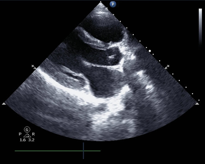
- Transesophageal echocardiography (TEE) is the reference standard for LAA thrombus detection.
- CCTA can be used as a routine diagnostic tool before and after LAA occlusion surgery.
Management
- Consider LLD-CCTA as an alternative to biphasic supine CCTA for LAA assessment.
Monitoring & Follow-up
- Follow-up with TEE within 48 hours after CCTA.
Risks
- Increased radiation exposure with conventional biphasic supine CCTA.
Patient & Prescribing Data
114 patients with nonvalvular atrial fibrillation.
LLD-CCTA maintains diagnostic accuracy while reducing radiation exposure.
Clinical Best Practices
- Utilize LLD positioning during CCTA to enhance LAA opacification.
- Adhere to ethical guidelines and obtain informed consent from patients.
Related Resources & Content