MRI-guided laser interstitial thermal therapy in epilepsy: indications, technique and outcome in an adult population. A single-center data analysis - Scorecard - MDSpire
Advertisement
MRI-guided laser interstitial thermal therapy in epilepsy: indications, technique and outcome in an adult population. A single-center data analysis
Clinical Scorecard: Laser Interstitial Thermal Therapy Guided by MRI for Epilepsy: Techniques, Indications, and Outcomes in Adults Based on a Single-Center Study
At a Glance
Category
Detail
Condition
Drug-resistant focal epilepsy
Key Mechanisms
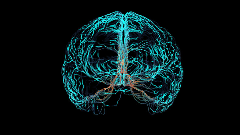
MRI-guided laser ablation using thermal energy to destroy epileptogenic tissue with real-time MRI thermometry monitoring
Target Population
Adults with drug-resistant focal epilepsy, including temporal lobe epilepsy with hippocampal sclerosis, hypothalamic hamartoma, focal cortical dysplasia, and periventricular heterotopia
Care Setting
Specialized epilepsy centers with multidisciplinary presurgical evaluation and MRIgLITT surgical capability
Key Highlights
MRIgLITT enables precise, minimally invasive ablation of epileptogenic lesions with millimeter accuracy and real-time thermal monitoring.
Indications include well-localized epileptogenic foci confirmed by concordant semiology, EEG, MRI, cognitive and functional studies.
Advantages include avoidance of craniotomy, treatment of deep or eloquent area lesions, rapid recovery, and cognitive function preservation.
Guideline-Based Recommendations
Diagnosis
Comprehensive presurgical evaluation including 3T MRI, video EEG, neuropsychological testing, and additional imaging (SPECT, PET, fMRI) as needed.
Multidisciplinary committee review to confirm concordance of seizure semiology, EEG onset, MRI findings, and cognitive profile before MRIgLITT indication.
Management
Use MRIgLITT primarily for dominant hemisphere temporal lobe epilepsy with hippocampal sclerosis to minimize cognitive impairment.
Select MRIgLITT as first-line surgical treatment for hypothalamic hamartoma and for lesions difficult to access by open surgery.
Standardize surgical protocols tailored to pathology (HS, HH, FCD, PVH, tumors) including catheter type, target and safety points.
Monitoring & Follow-up
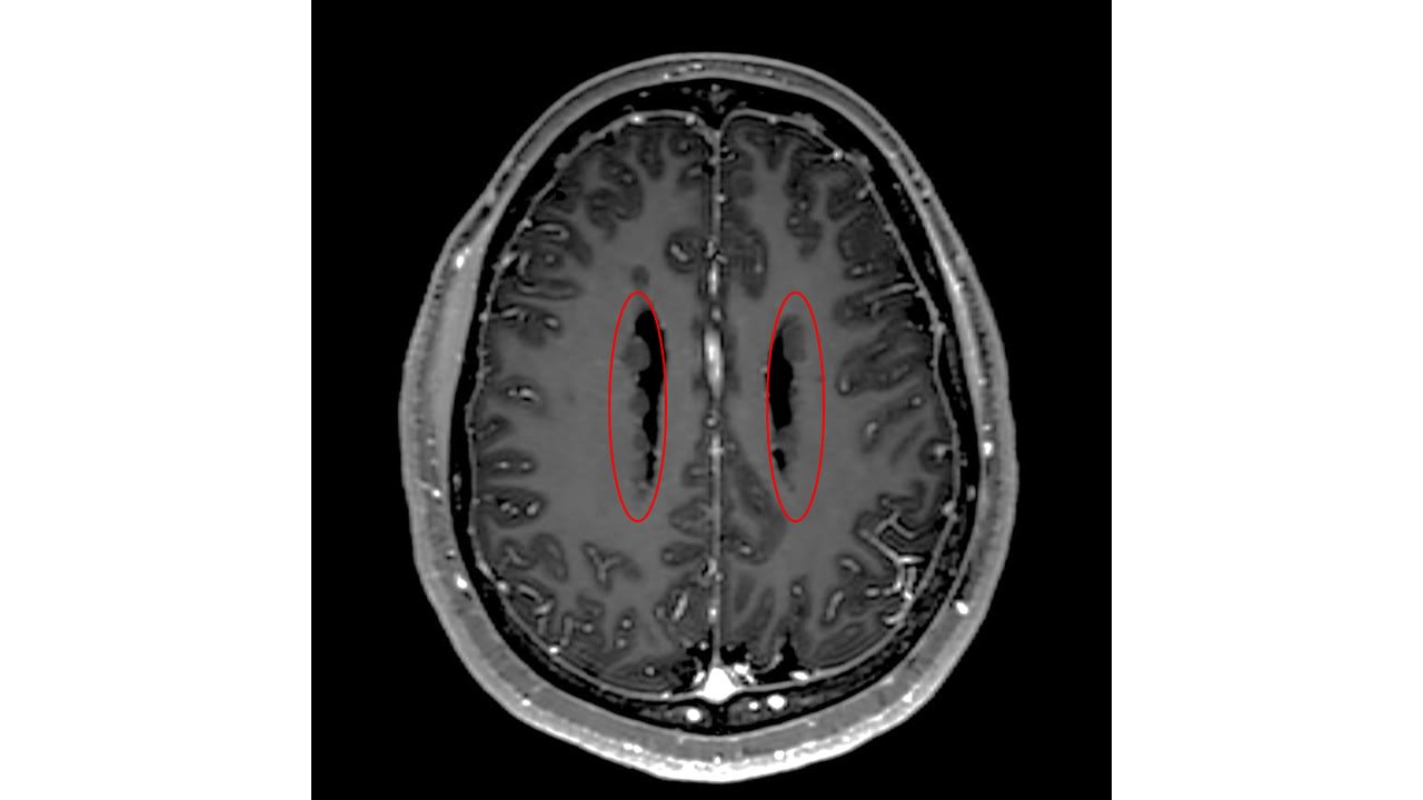
Continuous intraoperative MRI thermometry to monitor temperature and estimate irreversible tissue damage.
Postoperative seizure outcomes assessed by Engel Scale at multiple time points (3 months, 6 months, 1 year, 3 years).
Neuropsychological reassessment at 3 and 12 months post-surgery using Reliable Change Index to detect cognitive changes.
Risks
Potential complications related to thermal damage near critical structures; safety points and trajectory planning essential to minimize risks.
Unclear clinico-radiological predictors of outcomes necessitate further standardization and prospective studies.
Patient & Prescribing Data
Adults with drug-resistant focal epilepsy undergoing MRIgLITT at a single epilepsy center
MRIgLITT is effective for seizure control with cognitive preservation, especially in dominant temporal lobe epilepsy and hypothalamic hamartoma; treatment decisions guided by multidisciplinary evaluation and tailored surgical protocols.
Clinical Best Practices
Perform thorough multidisciplinary presurgical evaluation to ensure concordant localization of epileptogenic focus.
Customize MRIgLITT protocols by epilepsy pathology to optimize catheter placement, ablation parameters, and safety margins.
Use real-time MRI thermometry and software modeling to precisely control ablation extent and protect adjacent eloquent tissue.
Implement standardized neuropsychological assessments pre- and post-operatively to monitor cognitive outcomes.
Reserve MRIgLITT for lesions inaccessible or high-risk for open surgery and for patients prioritizing minimally invasive options.
by Nazaret Infante, Gerardo Conesa, Carmen Pérez-Enríquez, Jaume Capellades, Luísa Panadés de Oliveira, Laura Vilella, Alessandro Principe, Maria del Mar Crespi-Vallespir, Mireia Gallardo-Mir, Rodrigo Rocamora
Epilepsy remains a life-altering condition, particularly due to the unpredictable nature of seizures and their cumulative impact on cognition, independence and quality of life.