Clinical Scorecard: MRI-guided training approach for closure of the left atrial appendage
At a Glance
Category
Detail
Condition
Atrial fibrillation with risk of stroke due to left atrial appendage thrombus
Key Mechanisms
Percutaneous left atrial appendage (LAA) closure via transseptal puncture guided by patient-specific anatomical imaging
Target Population
Patients with atrial fibrillation undergoing LAA closure procedures
Care Setting
Interventional cardiology suites with imaging and 3D printing capabilities
Key Highlights
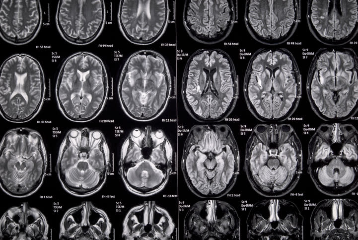
MRI-based 3D imaging enables patient-specific segmentation of left and right atria, LAA, and vena cava for procedural planning.
3D printed base model with predetermined transseptal puncture (TSP) sites and interchangeable silicone LAA models facilitates training and procedural simulation.
Non-contrast-enhanced respiratory navigated 3D isotropic MRI provides sufficient anatomical detail for segmentation without radiation or contrast exposure.
Guideline-Based Recommendations
Diagnosis
Use non-contrast-enhanced respiratory navigated 3D MRI to acquire patient-specific atrial and LAA anatomy prior to LAA closure.
Management
Plan transseptal puncture site based on patient-specific LAA anatomy to optimize sheath delivery and minimize multiple punctures.
Utilize 3D printed models for preprocedural planning and interventionalist training to improve procedural success.
Monitoring & Follow-up
Evaluate anatomical fit and sheath trajectory using 3D printed models before the procedure to reduce intra-procedural complications.
Risks
Repeated device implantation or multiple transseptal punctures may occur if patient-specific anatomy is not adequately assessed.
Radiation and contrast exposure risks are minimized by using MRI instead of CT for anatomical imaging.
Patient & Prescribing Data
Patients undergoing percutaneous LAA closure for stroke prevention in atrial fibrillation
MRI-based anatomical assessment and 3D printed training models support individualized device sizing and optimal puncture site selection, potentially reducing procedural complications.
Clinical Best Practices
Segment left and right atria, LAA, and vena cava from high-resolution MRI datasets for accurate anatomical modeling.
Incorporate multiple predetermined transseptal puncture sites in training models to simulate various clinical scenarios.
Use silicone models with tissue-mimicking elasticity to replicate LAA properties for realistic device deployment training.
Employ fused filament fabrication 3D printing with PLA filament for accessible and reproducible model production.
Leverage anatomical landmarks such as vena cava inferior and aortic root to guide puncture site placement.