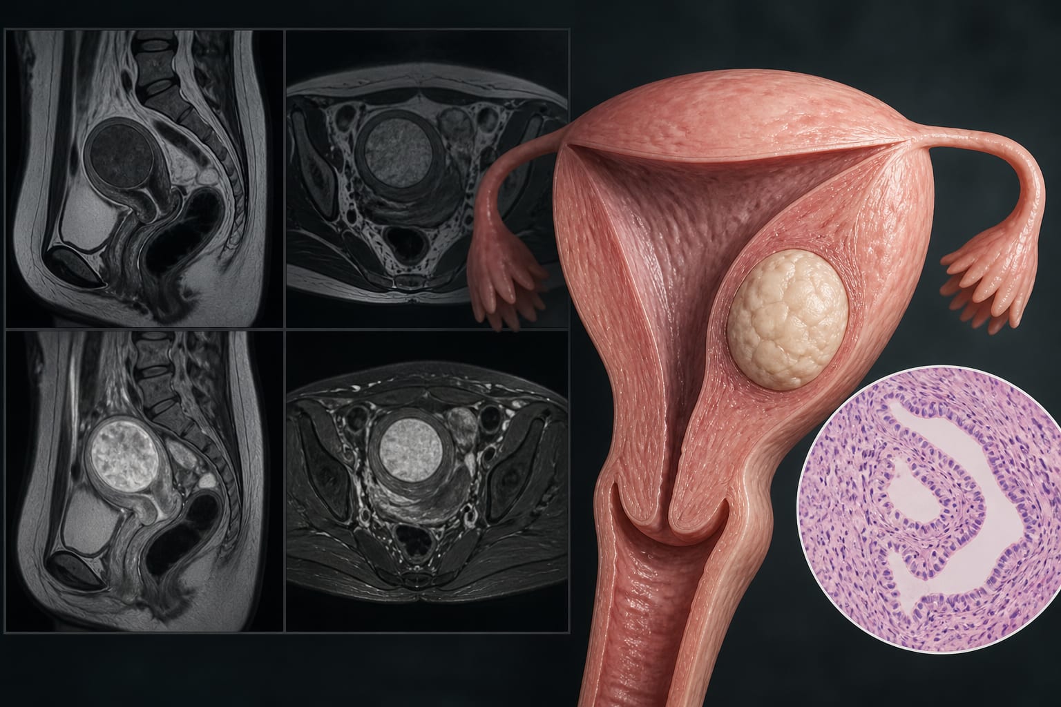
Magnetic Resonance Imaging Characteristics of Uterine Adenomatoid Tumors: A Study of Clinical and Histopathological Correlations
-
By
-
Changhuan Chen
-
Yudian Huang
-
Ping Chen
-
Caixia Zheng
-
Chengsheng Wang
-
Mingsi Yang
-
September 8, 2026
Clinical Scorecard: Magnetic Resonance Imaging Characteristics of Uterine Adenomatoid Tumors: A Study of Clinical and Histopathological Correlations
At a Glance
| Category | Detail |
|---|
| Condition | Uterine Adenomatoid Tumors |
| Key Mechanisms | Benign neoplasm arising from mesothelial tissue |
| Target Population | Women with uterine tumors |
| Care Setting | Clinical imaging and pathology |
Key Highlights
- Study analyzed 16 surgically resected uterine adenomatoid tumors
- MRI findings correlated with histopathologic features
- Lesions typically appear as solid masses without a discernible capsule
- Cystic degeneration and 'target sign' may be present on imaging
- Histopathologic examination is necessary for definitive diagnosis
Guideline-Based Recommendations
Diagnosis
- MRI is essential for confirming diagnosis and defining lesion extent
- Histopathologic examination is required for definitive diagnosis
Management
- Surgical treatment includes local excision or hysterectomy
Monitoring & Follow-up
- Regular imaging may be necessary to assess for changes in tumor characteristics
Risks
- Misdiagnosis as uterine leiomyomas may lead to inappropriate medical management
Patient & Prescribing Data
Women diagnosed with uterine adenomatoid tumors
Surgical intervention is the mainstay of treatment
Clinical Best Practices
- Utilize MRI for accurate preoperative assessment of uterine tumors
- Correlate imaging findings with histopathological data for diagnosis
- Be aware of the potential for misdiagnosis with uterine leiomyomas
Related Resources & Content