Minimally invasive resection of solid intraventricular lesions via single burr-hole ventriculoscopy
-
By
-
Xinghua Xu
-
Jiashu Zhang
-
Zhichao Gan
-
Qun Wang
-
Haoyang Zheng
-
Shiyu Zhang
-
Xiaolei Chen
-
June 15, 2026
-
Clinical Scorecard: Single Burr-Hole Ventriculoscopy for the Minimally Invasive Resection of Solid Intraventricular Lesions
At a Glance
| Category | Detail |
|---|
| Condition | Solid intraventricular lesions |
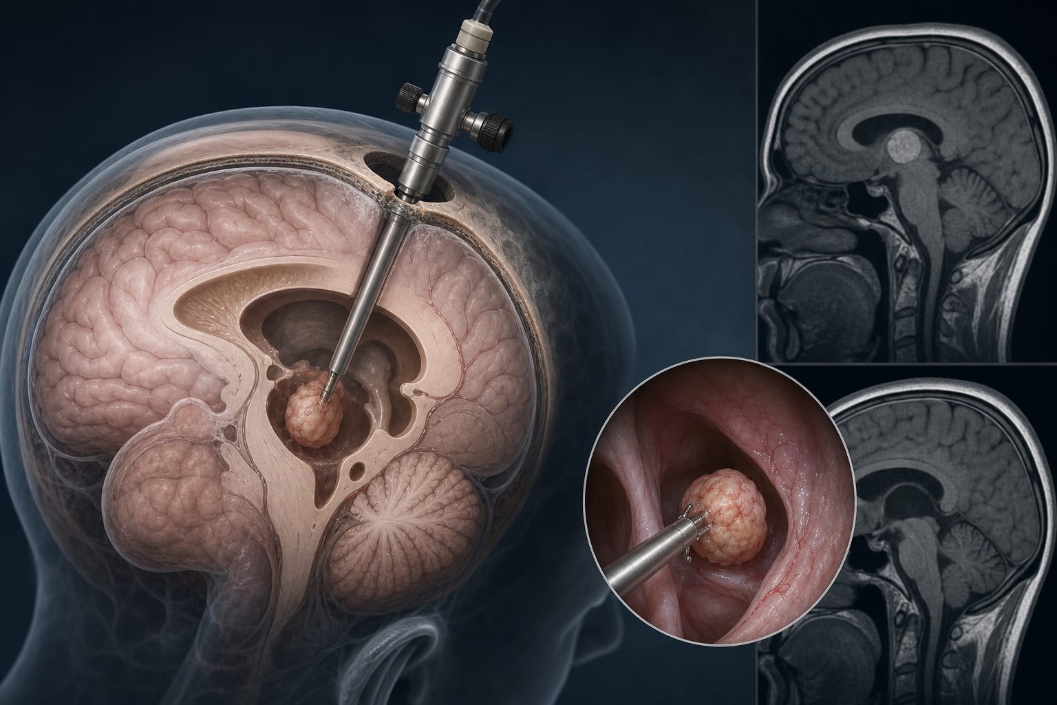
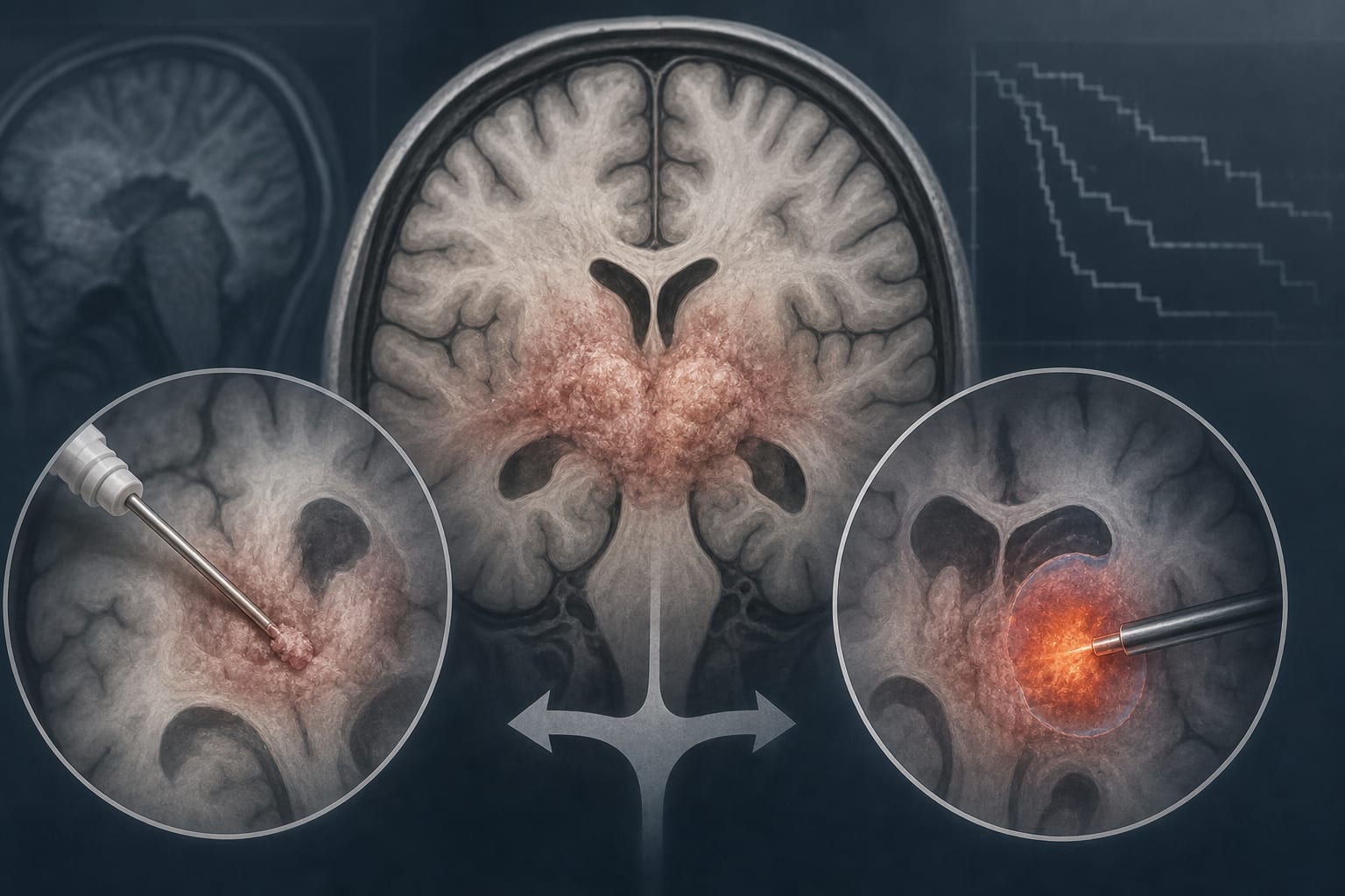
| Key Mechanisms | Single burr-hole pure ventriculoscopic resection |
| Target Population | Patients with solid intraventricular lesions |
| Care Setting | Neurosurgical department |
Key Highlights
- Gross-total resection achieved in 92.1% of cases
- No seizures, cerebrospinal fluid leakage, or permanent neurological deficits reported
- Transient diplopia in 5.3% of patients; one intracranial infection in 2.6%
- Mean follow-up of 60 months with no definite lesion recurrence
- High-resolution 3D-SPACE MRI used for individualized trajectory planning
Guideline-Based Recommendations
Diagnosis
- Use high-resolution 3D-SPACE MRI for imaging solid intraventricular lesions
Management
- Consider single burr-hole pure ventriculoscopic resection for selected small solid intraventricular lesions
Monitoring & Follow-up
- Postoperative MRI to evaluate extent of resection and follow-up for neurological outcomes
Risks
- Potential for transient diplopia, intracranial infection, and delayed hydrocephalus
Patient & Prescribing Data
Patients with radiologically confirmed solid intraventricular lesions
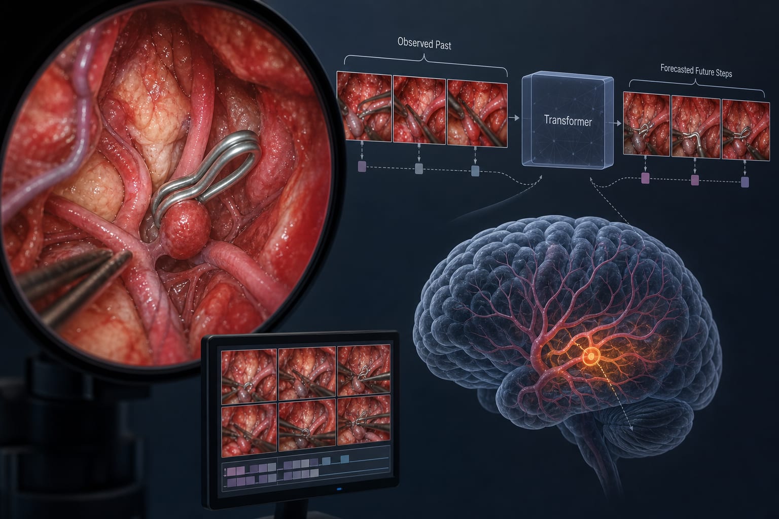
Surgical approach minimizes invasiveness and preserves surrounding neural structures
Clinical Best Practices
- Utilize virtual ventriculoscopy for preoperative planning
- Ensure careful selection of patients based on lesion characteristics and anatomical considerations
Related Resources & Content