Surgery assistance system for continuous resection of brain tumors-proposal of continuous tumor resection forceps, tumor cell separation, dehydration, and isolation mechanism - Scorecard - MDSpire
Advertisement
Surgery assistance system for continuous resection of brain tumors-proposal of continuous tumor resection forceps, tumor cell separation, dehydration, and isolation mechanism
Clinical Scorecard: Innovative Surgical Assistance System for Continuous Resection of Brain Tumors: Introduction of Continuous Tumor Resection Forceps and Mechanisms for Tumor Cell Separation, Dehydration, and Isolation
At a Glance
Category
Detail
Condition
Malignant brain tumors with unclear boundaries between tumor and normal brain tissue
Key Mechanisms
Continuous tumor resection forceps with suction and reflux cleaning water, separation and dehydration mechanism to remove excess fluid, tumor cell isolation, and flow cytometer-based malignancy diagnosis
Target Population
Patients undergoing brain tumor surgery requiring maximal tumor resection while preserving normal brain tissue
Care Setting
Intraoperative neurosurgical setting with integrated surgical navigation and rapid pathological diagnosis
Continuous tumor resection forceps enable continuous tumor removal with integrated suction and saline reflux cleaning.
Separation and dehydration mechanisms reduce dilution of tumor cells for accurate flow cytometry-based intraoperative malignancy diagnosis.
Guideline-Based Recommendations
Diagnosis
Use intraoperative rapid diagnosis with pathological specimens obtained via biopsy to determine tumor malignancy.
Employ flow cytometry with fluorescent DNA staining for fast malignancy diagnosis of isolated tumor cells.
Management
Maximize tumor resection ratio while preserving normal brain tissue to reduce recurrence and improve survival.
Utilize continuous tumor resection forceps to facilitate continuous tumor removal and increase specimen number.
Incorporate surgical navigation systems integrating neural pathways and vascular structures to guide resection.
Monitoring & Follow-up
Monitor tumor cell concentration during intraoperative diagnosis to avoid dilution effects from reflux cleaning water.
Display diagnostic results on navigation system to guide extent of tumor extraction.
Risks
Avoid injury to normal brain tissue to prevent sequelae such as speech disorders or hemiplegia.
Minimize tumor cell leakage by ensuring adequate sampling and precise excision range determination.
Patient & Prescribing Data
Patients undergoing surgical resection of malignant brain tumors
Higher tumor resection ratios (≥90-95%) correlate with improved 5-year survival; continuous resection and rapid diagnosis support maximizing resection while minimizing normal tissue damage.
Clinical Best Practices
Determine exact tumor extraction range intraoperatively using rapid pathological diagnosis to balance maximal tumor removal and normal tissue preservation.
Use continuous tumor resection forceps with suction and saline reflux to enable continuous tumor removal and reduce surgeon burden.
Implement separation and dehydration mechanisms to prepare tumor cells for accurate flow cytometry diagnosis.
Integrate intraoperative navigation systems with neural and vascular pathway mapping to improve surgical precision.
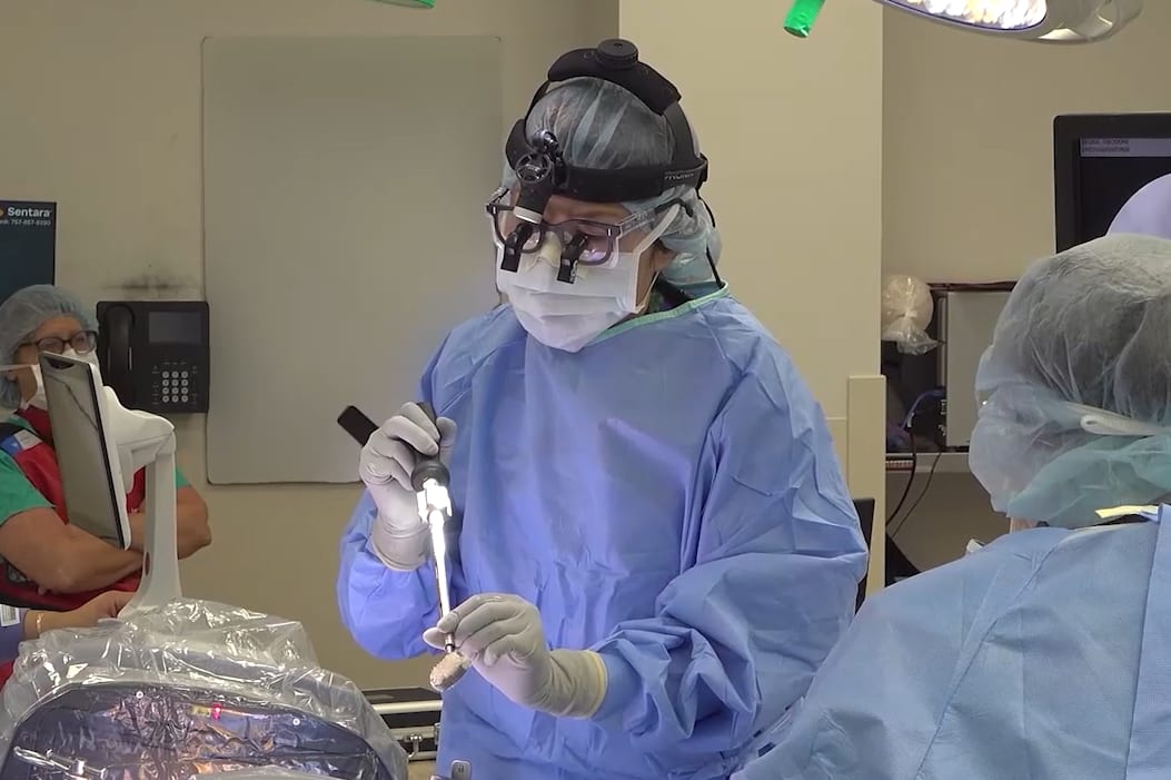
In this procedural case review, vascular surgeon Dr. Samuel Steerman and neurosurgeon Dr. Shannon Clark collaborate to perform an anterior lumbar interbody fusion (ALIF).