A case report and literature review on imaging manifestations of immature teratoma in fetal oral cavity with concurrent intracranial abnormalities - Scorecard - MDSpire
Advertisement
A case report and literature review on imaging manifestations of immature teratoma in fetal oral cavity with concurrent intracranial abnormalities
Clinical Scorecard: Imaging Features of Fetal Oral Cavity Immature Teratoma with Associated Intracranial Abnormalities: A Case Study and Literature Review
At a Glance
Category
Detail
Condition
Key Mechanisms
Abnormal proliferation of germ cells leading to teratoma formation, with potential for concurrent intracranial abnormalities such as arachnoid cysts and meningoceles.
Target Population
Care Setting
Key Highlights
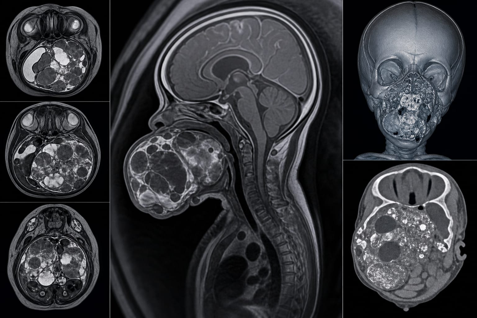
Large solid-cystic mass in the fetal oropharynx associated with maxilla and skull base.
CT imaging shows heterogeneous density and punctate calcifications.
MRI reveals irregular mass with specific signal characteristics indicating immature neuroepithelial elements.
Concurrent intracranial abnormalities include arachnoid cyst and meningocele.
Early diagnosis is crucial due to high morbidity and mortality risks.
Guideline-Based Recommendations
Diagnosis
Management
Consider termination of pregnancy in cases of severe fetal abnormalities.
Explore options for monitoring and supportive care in less severe cases.
Monitoring & Follow-up
Risks
Patient & Prescribing Data
Induction of labor may be indicated in cases of severe fetal abnormalities, but consider other management strategies based on individual cases.
Clinical Best Practices
Conduct thorough imaging evaluations to differentiate between immature and mature teratomas.
Document imaging findings systematically to enhance diagnostic accuracy.
Encourage interdisciplinary collaboration for comprehensive management.