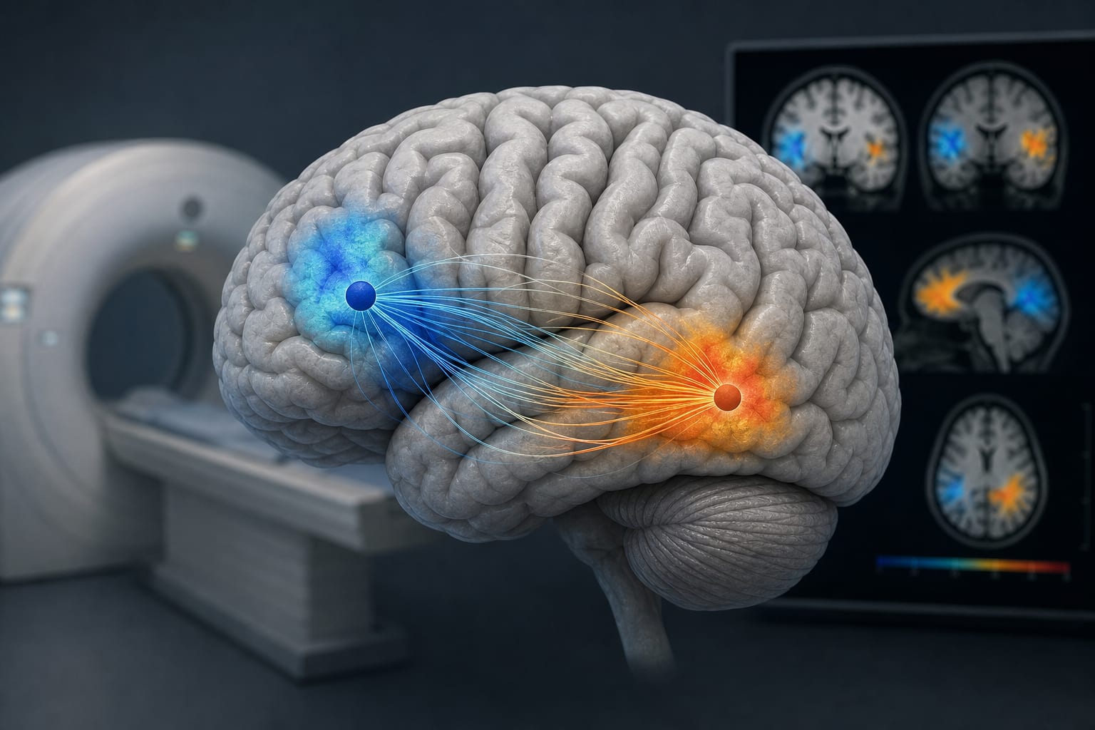
Application Value of Resting-State fMRI in Preoperative Lateralization of Language Areas in Epilepsy with Left-Sided Epileptogenic Foci
By
Wang, Zihao
Hu, Tian-Qi
Meng, Wang
Li, Ying
Sun, Yaning
Zhang, Di
Li, Wen-Ling
May 29, 2026
Clinical Scorecard: Utilization of Resting-State fMRI for Preoperative Language Area Lateralization in Patients with Left-Sided Epileptogenic Foci
At a Glance
Category Detail
Condition Left-sided Epileptogenic Foci in Epilepsy Key Mechanisms Language lateralization differences and functional connectivity analysis using resting-state fMRI. Target Population Patients with left-sided epileptogenic foci and healthy controls. Care Setting Preoperative assessment in epilepsy surgery.
Key Highlights
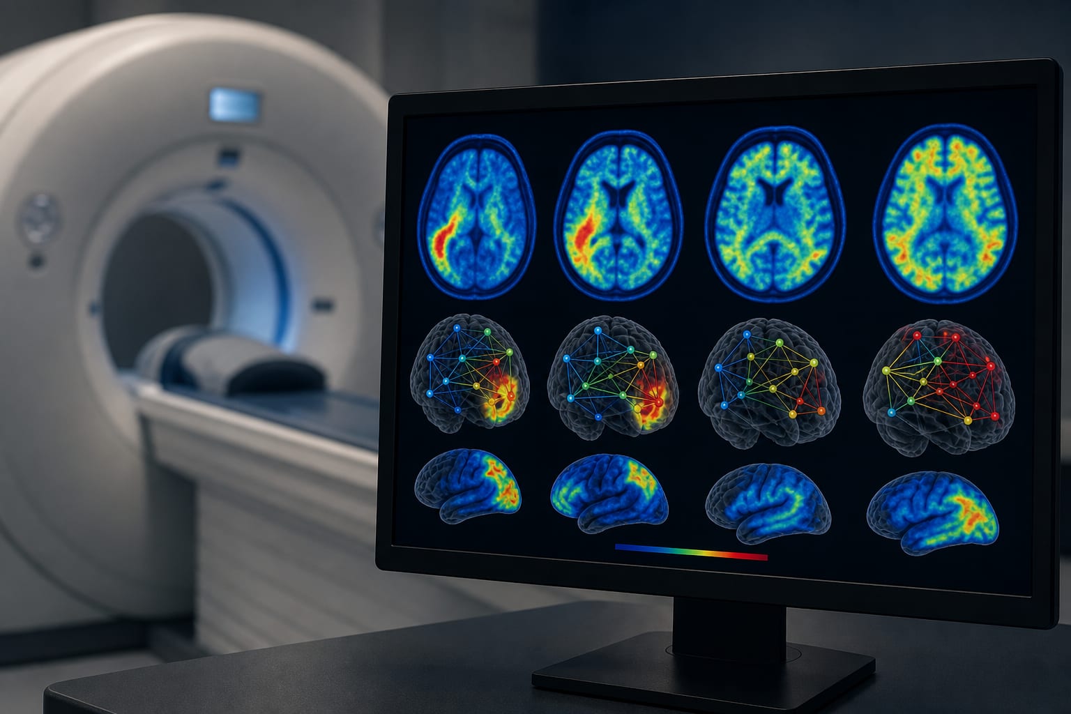
Non-classical language dominance observed in 69.4% of epilepsy patients compared to 45.5% in controls. Wernicke's area showed a higher shift in lateralization compared to Broca's area. The combined laterality index–activation map scheme demonstrated 83.3% consistency. Guideline-Based Recommendations
Diagnosis
Utilize resting-state fMRI to assess language lateralization in epilepsy patients. Management
Prioritize protection of the left Broca's area during epilepsy surgeries involving the left frontotemporal lobe. Monitoring & Follow-up
Conduct SEEG cortical stimulation and postoperative follow-up to verify language function. Risks
Increased risk of language impairment associated with resection of the left Wernicke's area. Patient & Prescribing Data
36 patients with left-sided epileptogenic foci.
Surgical candidates should undergo SEEG stimulation to assess language function reliability.
Clinical Best Practices
Employ the laterality index–activation map scheme for preoperative language assessment. Monitor language function closely post-surgery, especially after left frontal lobe resections. Related Resources & Content