Quantifying knee cartilage development trajectories in children aged 6–12 years via diffusion tensor imaging
-
By
-
Zhuo Cheng
-
Wei Li
-
Wei Ma
-
Gaohui Zhu
-
Yujuan Hu
-
Junya Ma
-
Sijie Gao
-
Yilu Zhang
-
Hailun Peng
-
Ye Xu
-
July 1, 2026
-
Clinical Scorecard: Assessing Pediatric Knee Cartilage Growth Patterns in Children Aged 6 to 12 Years Using Diffusion Tensor Imaging
At a Glance
| Category | Detail |
|---|
| Condition | Pediatric Knee Cartilage Development |
| Key Mechanisms | Diffusion Tensor Imaging (DTI) biomarkers: Fractional Anisotropy (FA) and Apparent Diffusion Coefficient (ADC) |
| Target Population | Healthy children aged 6–12 years |
| Care Setting | Pediatric radiology and imaging |
Key Highlights
- FA values increased significantly with age and bone age, correlating best with bone age.
- Girls exhibited higher FA values than boys across all age groups.
- ADC values in the growth plate decreased with increasing bone age, with a steeper decline in girls.
- Excellent reproducibility of DTI measurements was confirmed (FA/ADC ICC > 0.94).
- DTI biomarkers sensitively reflect pediatric knee cartilage maturation.
Guideline-Based Recommendations
Diagnosis
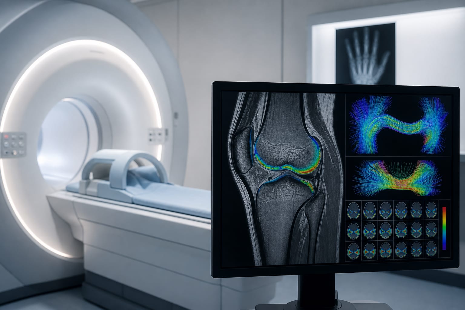
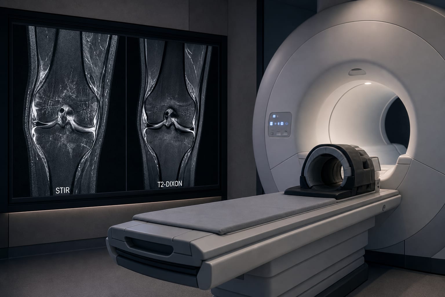
- Utilize DTI to assess cartilage microstructure and development.
Management
- Monitor cartilage development and pathology in children using DTI biomarkers.
Monitoring & Follow-up
- Regularly evaluate FA and ADC values to track cartilage maturation.
Risks
- Early detection of pathological changes is crucial to prevent growth retardation and skeletal deformities.
Patient & Prescribing Data
Healthy children aged 6–12 years without joint abnormalities.
No specific treatments are prescribed; focus on monitoring cartilage development.
Clinical Best Practices
- Conduct comprehensive physical examinations to exclude joint abnormalities.
- Use standardized methods for bone age assessment.
- Ensure reproducibility of DTI measurements through careful imaging protocols.
Related Resources & Content