Clinical Scorecard: The Relationship Between Shunt Dependency and Tumor Resection Extent in Supratentorial Intraventricular Tumors
At a Glance
Category
Detail
Condition
Supratentorial intraventricular tumors (SIVTs)
Key Mechanisms
Tumor mass effect causing obstructive hydrocephalus due to blockage of cerebrospinal fluid flow within the ventricular system
Target Population
Patients with tumors located at least partially in the supratentorial ventricular system (lateral ventricles and third ventricle)
Care Setting
Academic neuro-oncology centers with neurosurgical and neuro-oncological expertise
Key Highlights
SIVTs are rare, comprising 1–3% of intracranial lesions, with diverse histological entities arising primarily or secondarily within the ventricular system.
Surgical management aims for maximal safe tumor resection via various approaches tailored to tumor location, balancing tumor removal and preservation of eloquent brain structures.
Multiple shunting strategies exist to manage obstructive hydrocephalus perioperatively, including external ventricular drainage, ventriculoperitoneal/atrial shunts, endoscopic third ventriculostomy, and stereotactic internal shunting.
Guideline-Based Recommendations
Diagnosis
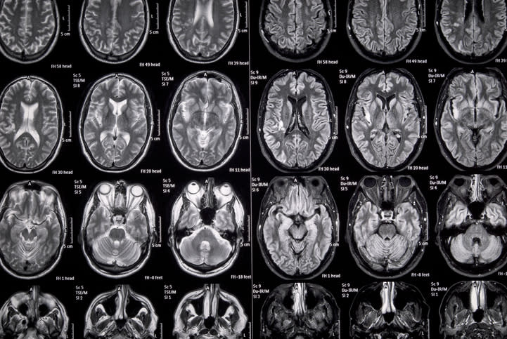
Use MRI with contrast-enhanced T1-weighted and FLAIR/T2 sequences for tumor volume assessment and hydrocephalus evaluation.
Diagnose occlusive hydrocephalus radiologically by ventricular enlargement and transependymal CSF diapedesis.
Histopathological diagnosis should follow the 2021 WHO CNS tumor classification based on tissue obtained via microsurgical resection or biopsy.
Management
First-line therapy for symptomatic SIVTs is complete microsurgical or neuroendoscopic tumor resection.
Adjuvant therapy is considered depending on histological tumor entity.
Benign cystic lesions (e.g., colloid or pineal cysts) may be managed with minimally invasive stereotactic procedures.
Shunting strategies for hydrocephalus include perioperative external ventricular drainage, ventriculoperitoneal or atrial shunts, endoscopic third ventriculostomy, and stereotactic internal shunting.
Choice of shunting procedure should be individualized; the necessity of shunting after complete tumor removal remains unclear.
Monitoring & Follow-up
Follow-up imaging per institutional guidelines based on tumor type and clinical status.
Surveillance MRI to assess for residual tumor and hydrocephalus.
Monitor for new or worsening neurological deficits postoperatively.
Risks
Surgical risks include injury to eloquent deep brain and neurovascular structures.
Postoperative neurological deficits may range from mild to severe, including seizures, cranial nerve deficits, aphasia, motor/sensory impairments, and cognitive decline.
Hydrocephalus persistence may require additional shunting procedures.
Patient & Prescribing Data
Patients undergoing microsurgical or neuroendoscopic resection for supratentorial intraventricular tumors with or without hydrocephalus
Extent of tumor resection and choice of shunting strategy influence postoperative hydrocephalus management; individualized treatment plans are essential.
Clinical Best Practices
Perform maximal safe tumor resection tailored to tumor location within the ventricular system to minimize brain retraction and preserve critical structures.
Use volumetric MRI analysis to define extent of resection and residual tumor burden.
Assess and manage hydrocephalus perioperatively with appropriate shunting techniques based on clinical and imaging findings.
Engage interdisciplinary brain tumor boards for diagnostic and therapeutic decision-making.
Classify postoperative neurological deficits by severity to guide management and rehabilitation.
by Nico Teske, Mariana Chiquillo-Domínguez, Benjamin Skrap, Patrick N. Harter, Kai Rejeski, Jens Blobner, Louisa von Baumgarten, Joerg-Christian Tonn, Mathias Kunz, Niklas Thon, Philipp Karschnia