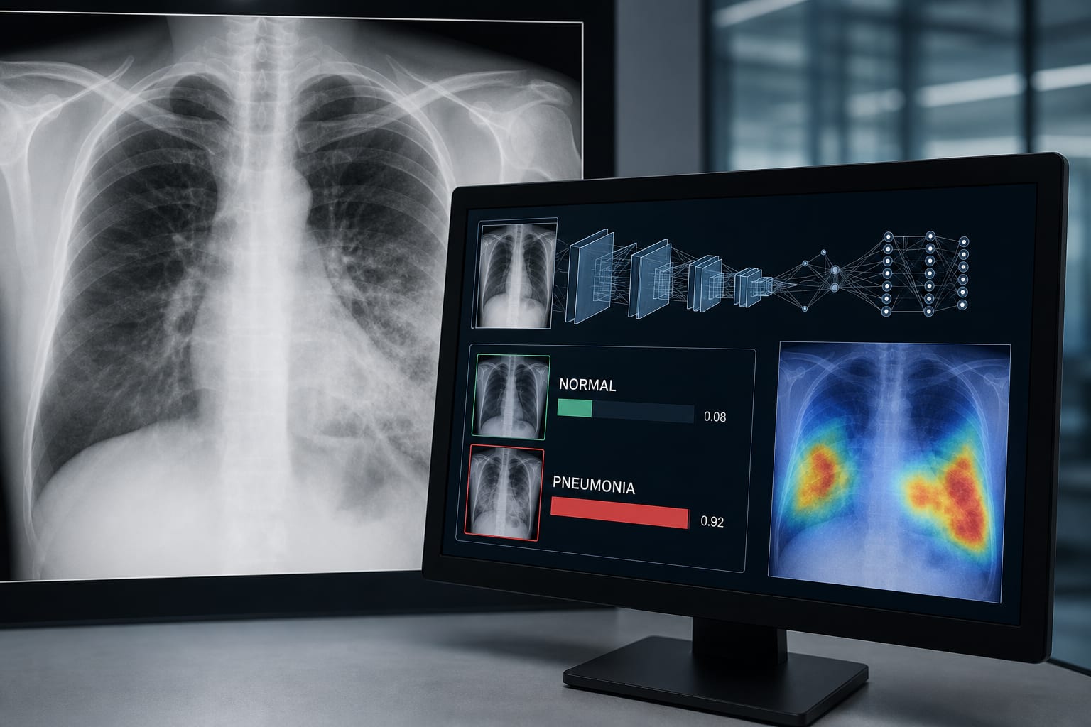
Application of deep learning methods in the classification of normal and pneumonia lung images
-
By
-
Mehmet Kabak
-
Mehmet Tarık Baran
-
Barış Çil
-
May 21, 2026
Clinical Scorecard: Utilization of Deep Learning Techniques for Differentiating Normal Lung Images from Pneumonia in Chest Radiographs
At a Glance
| Category | Detail |
|---|
| Condition | Pneumonia |
| Key Mechanisms | Deep learning-based models for automated classification of chest X-ray images. |
| Target Population | Adults (≥ 18 years) with suspected pneumonia. |
| Care Setting | Tertiary hospital with a focus on chest radiography. |
Key Highlights
- VGG16 achieved the highest test accuracy (87.1%) and AUC (0.92).
- InceptionV3 performed well with 85.0% accuracy and AUC of 0.84.
- Xception model showed lower sensitivity in detecting pneumonia cases.
- ResNet50 underperformed with an accuracy of 74.2% and AUC of 0.81.
- Study emphasizes the need for larger, balanced datasets for improved sensitivity.
Guideline-Based Recommendations
Diagnosis
- Utilize chest X-rays for initial pneumonia diagnosis.
- Implement deep learning models to enhance diagnostic accuracy.
Management
- Consider automated classification systems as adjuncts to radiological evaluation.
Monitoring & Follow-up
- Regularly evaluate model performance using metrics such as accuracy and AUC.
Risks
- Be aware of potential class imbalance and overfitting in model training.
Patient & Prescribing Data
Adults with symptoms of pneumonia and diagnostic-quality PA chest radiographs.
Early diagnosis through automated systems can improve management outcomes.
Clinical Best Practices
- Use standardized PA chest radiographs for model training.
- Ensure high-quality imaging to avoid misclassification.
- Incorporate clinical context in radiological assessments.
Related Resources & Content