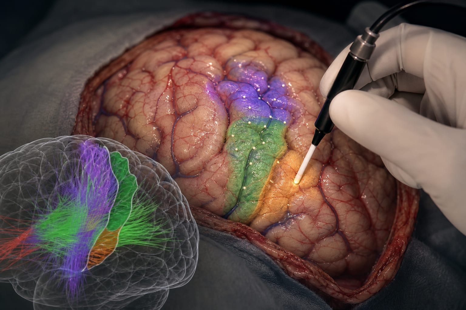
A Combined Exo- and Endoscopic Technique for Managing Recurrent Vestibular Schwannomas After Surgery and Radiosurgery: My Methodology
-
By
-
Kenichiro Iwami
-
Kazuhito Takeuchi
-
Yuichi Nagata
-
Ryuta Saito
-
August 5, 2025
Clinical Scorecard: A Combined Exo- and Endoscopic Technique for Managing Recurrent Vestibular Schwannomas After Surgery and Radiosurgery: My Methodology
At a Glance
| Category | Detail |
|---|---|
| Condition | Recurrent vestibular schwannomas (VS) after surgery and radiosurgery |
| Key Mechanisms | Surgical resection using a combined exoscopic and endoscopic two-step approach (EETA) to improve visualization and removal of intracanalicular and extracanalicular tumour components |
| Target Population | Patients with recurrent or residual vestibular schwannomas after initial surgery and stereotactic radiosurgery |
| Care Setting | Neurosurgical operating room with access to exoscope and rigid endoscopes |
Key Highlights
- The retrosigmoid approach (RSA) allows access to the cerebellopontine angle and opening of the internal auditory canal (IAC) but has limited visualization of the fundus of the IAC (FIAC).
- The exo- and endoscopic two-step approach (EETA) combines exoscopic removal of extracanalicular tumour and endoscopic resection of residual intracanalicular tumour, including use of a 70-degree endoscope to visualize all four FIAC quadrants.
- Salvage surgery with EETA facilitates meticulous observation and removal of tumour in the IAC and CPA, improving resection completeness and reducing recurrence risk.
Guideline-Based Recommendations
Diagnosis
- Use gadolinium-enhanced MRI to identify residual or recurrent vestibular schwannoma in the IAC and CPA.
- Assess tumour growth after initial surgery and radiosurgery to determine need for salvage intervention.
Management
- Perform salvage surgery via retrosigmoid approach with combined exoscopic and endoscopic visualization (EETA) for recurrent VS.
- Use microsurgical instruments under stereoscopic exoscopic visualization for extracanalicular tumour resection.
- Employ rigid endoscopes (0°, 30°, 70°) to inspect and remove residual intracanalicular tumour, especially at the FIAC.
- Consider further bone removal under endoscopy if FIAC exposure is inadequate.
- Use curved instruments to minimize bone removal from FIAC when possible.
Monitoring & Follow-up
- Postoperative MRI to confirm complete tumour resection and absence of residual tumour.
- Clinical monitoring for facial nerve function and hearing status post-surgery.
Risks
- Adhesions and capsule formation from prior treatments may complicate tumour dissection.
- Risk of facial nerve injury necessitates careful identification and preservation during surgery.
- Incomplete visualization of FIAC may lead to residual tumour and recurrence.
Patient & Prescribing Data
Patients with recurrent vestibular schwannomas after initial surgery and radiosurgery
Salvage surgery using EETA can be performed safely with no facial paralysis and effective tumour control demonstrated in reported cases.
Clinical Best Practices
- Position patient in semilateral position and reopen previous craniotomy site for retrosigmoid approach.
- Use exoscope for extracanalicular tumour removal with microsurgical instruments under stereoscopic visualization.
- Widen IAC opening as needed to access intracanalicular tumour components.
- Use 70-degree endoscope to visualize all four quadrants of the FIAC for thorough tumour resection.
- Perform additional bone removal under endoscopy if FIAC exposure is insufficient.
- Identify facial nerve early, especially when adhesions obscure anatomy, to prevent nerve injury.
References
- Use of endoscopic assistance in retrosigmoid approach for vestibular schwannomas
- Management of recurrent vestibular schwannomas with stereotactic radiosurgery
- Salvage surgery techniques for recurrent vestibular schwannomas
Based on findings from:
Exo- and endoscopic two-step approach for recurrent vestibular schwannomas following surgical resection and radiosurgery: How I do it
Kenichiro Iwami, Kazuhito Takeuchi, Yuichi Nagata, Ryuta Saito. Acta Neurochirurgica, 2025.
https://link.springer.com/article/10.1007/s00701-025-06625-1
This content is an AI-generated, fully rewritten summary based on a published scholarly article. It does not reproduce the original text and is not a substitute for the original publication. Readers are encouraged to consult the source for full context, data, and methodology.