Utilizing fully-automated 3D organ segmentation for hepatic steatosis assessment with CT attenuation-based parameters
-
By
-
Jeongin Yoo
-
Ijin Joo
-
Sun Kyung Jeon
-
Junghoan Park
-
Soon Ho Yoon
-
February 23, 2024
-
Clinical Scorecard: Employing Fully-Automated 3D Organ Segmentation for Assessing Hepatic Steatosis Using CT Attenuation Metrics
At a Glance
| Category | Detail |
|---|
| Condition | Hepatic steatosis (fat accumulation in hepatocytes) in non-alcoholic fatty liver disease |
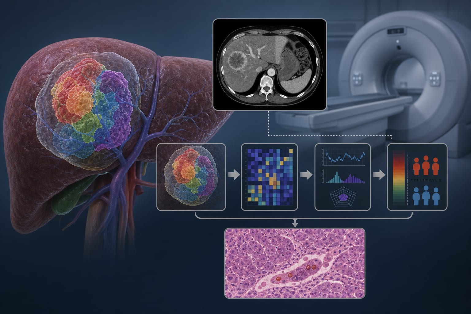
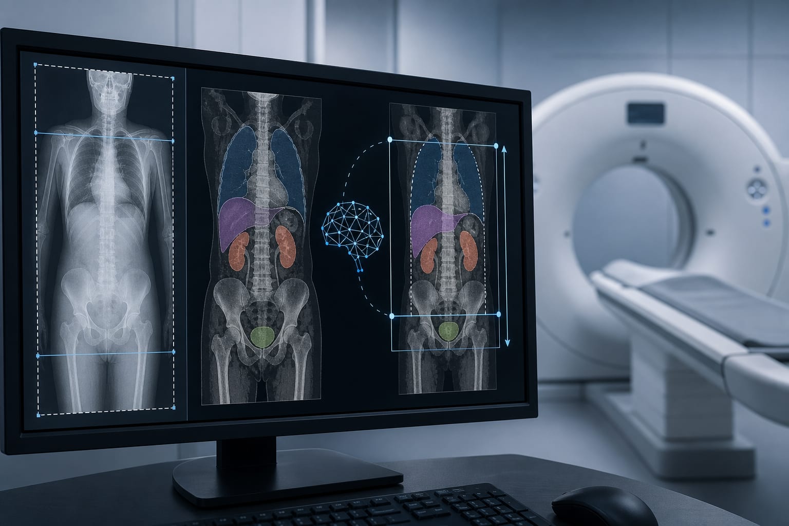
| Key Mechanisms | Fat accumulation decreases CT attenuation values of liver tissue; automated 3D segmentation enables volumetric CT attenuation measurement |
| Target Population | Adults undergoing preoperative liver evaluation, including potential living liver donors |
| Care Setting | Radiology departments performing CT and MRI imaging for hepatic steatosis assessment |
Key Highlights
- Liver biopsy is gold standard but invasive; non-invasive imaging (MRI-PDFF, MRS) preferred for hepatic steatosis quantification
- CT attenuation values inversely correlate with hepatic fat content; ROI-based CT measurements have moderate accuracy (AUC ~0.7–0.9)
- Fully automated 3D organ segmentation allows volumetric CT attenuation measurement, potentially improving assessment of uneven steatosis
Guideline-Based Recommendations
Diagnosis
- Use MRI-PDFF or MRS as non-invasive reference standards for hepatic steatosis quantification
- CT can be used opportunistically for hepatic steatosis screening, preferably with pre-contrast images
- Manual ROI-based CT attenuation measurements involve multiple liver regions and spleen for comparison
Management
- Employ non-invasive imaging techniques for routine and repeated hepatic steatosis monitoring
- Consider volumetric CT attenuation measurements via automated 3D segmentation to improve assessment accuracy
Monitoring & Follow-up
- Monitor hepatic steatosis progression or improvement using MRI-PDFF or MRS as reference
- CT attenuation values can be tracked over time, noting limitations in mild steatosis detection
Risks
- CT involves ionizing radiation exposure; use judiciously especially for repeated assessments
- Post-contrast CT can be used but pre-contrast CT preferred for attenuation measurements
- ROI-based CT measurements may be less representative in heterogeneous steatosis
Patient & Prescribing Data
Adults evaluated for hepatic steatosis, including potential living liver donors
Non-invasive imaging preferred over biopsy; automated volumetric CT attenuation measurement may enhance screening and monitoring
Clinical Best Practices
- Use MRI-PDFF or MRS as non-invasive gold standards for hepatic fat quantification
- Perform CT attenuation measurements on pre-contrast images when possible
- Apply fully automated 3D organ segmentation for volumetric liver and spleen attenuation measurements to improve accuracy
- Place multiple ROIs in different liver segments and spleen for manual CT attenuation assessment
- Avoid large vessels, bile ducts, and liver edges when positioning spectroscopy voxels or ROIs
References