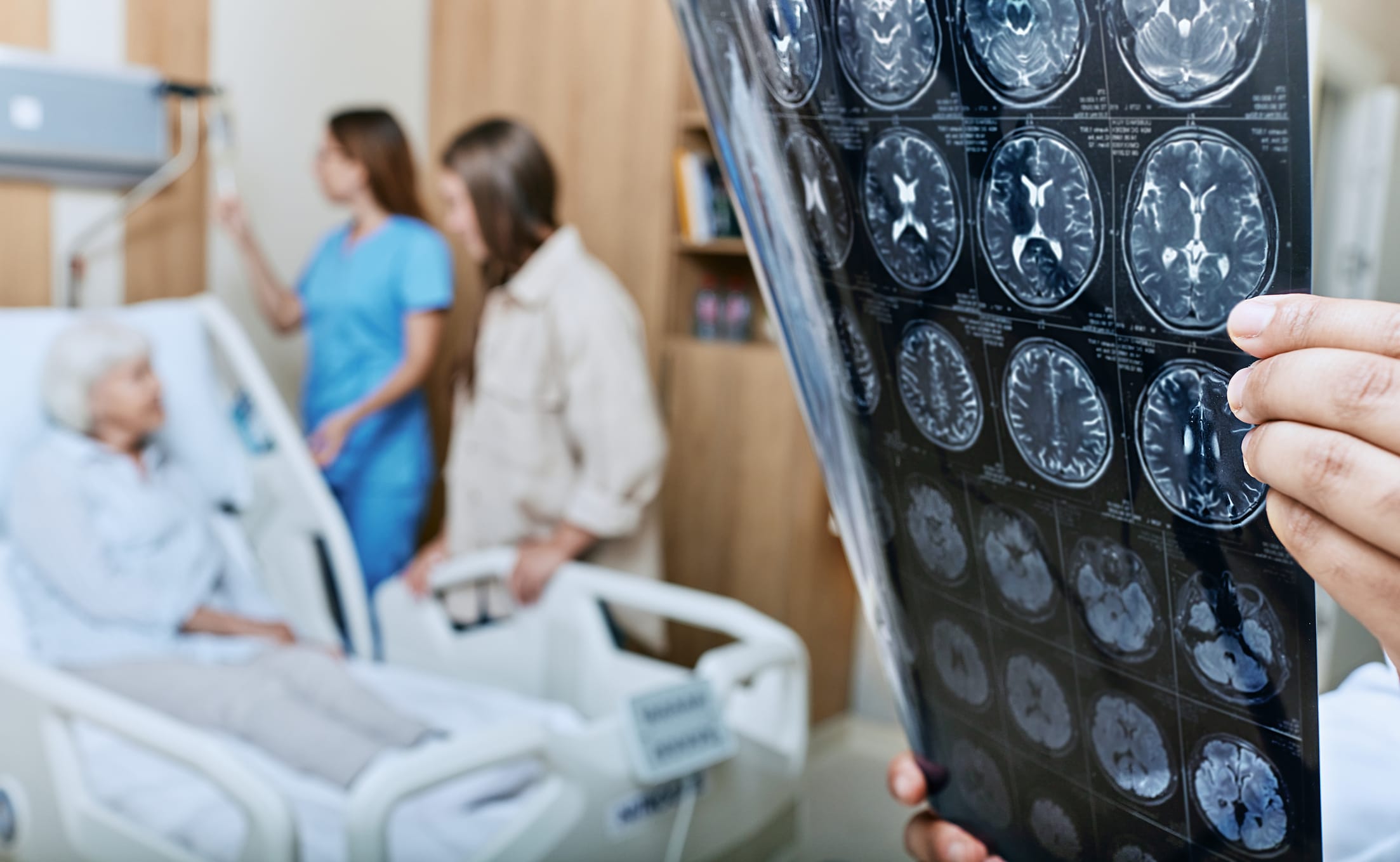
Patient positioning in lounging or (semi-)sitting position affects cerebral blood flow and risk of venous air embolism (VAE)
Target Population
Patients undergoing neurosurgery in posterior fossa region
Care Setting
Operating room during neurosurgical procedures
Key Highlights
Lounging and (semi-)sitting positions facilitate better surgical access and cranial nerve preservation in posterior fossa surgery.
Venous air embolism (VAE) is a critical risk due to hydrostatic pressure differences; severity depends on air volume per time entering circulation.
Transesophageal echocardiography (TEE) is essential for early detection and grading of VAE, guiding targeted intraoperative management.
Guideline-Based Recommendations
Diagnosis
Assess for persistent foramen ovale (PFO) preoperatively to evaluate risk of paradoxical embolism.
Use transesophageal echocardiography (TEE) intraoperatively to detect and grade venous air embolism (VAE) using scales such as the Tuebingen Venous Air Embolism Grading Scale.
Management
Communicate promptly with the surgical team upon VAE detection to prevent further air entry.
Treat hemodynamic depression caused by VAE with appropriate supportive measures.
Consider aspiration of air or 'air lock' if indicated by severity of VAE.
Monitoring & Follow-up
Vigilant perioperative anesthesiological monitoring to ensure adequate cerebral blood flow.
Continuous hemodynamic monitoring and intraoperative ventilation adjustments tailored to patient positioning.
Risks
Risk of venous air embolism due to elevated surgical site relative to heart.
Potential for paradoxical embolism in patients with PFO leading to stroke or myocardial infarction.
Physiological changes induced by sitting position including hemodynamic alterations.
Patient & Prescribing Data
Patients undergoing neurosurgical procedures in lounging, sitting, or semi-sitting positions, especially those with posterior fossa pathology.
Anesthetic management must focus on early detection and treatment of VAE, maintenance of cerebral perfusion, and careful positioning to minimize risks.
Clinical Best Practices
Preoperative screening for PFO to stratify embolism risk.
Use of TEE for real-time detection and grading of VAE during surgery.
Close interdisciplinary communication between anesthesiologists and surgeons for timely intervention.
Careful positioning with attention to cervical spine flexion and rotation to optimize surgical access while minimizing complications.
Continuous hemodynamic and respiratory monitoring tailored to the physiological changes of the sitting position.