Clinical Scorecard: Individualized Treatment Approaches for Diabetic Macular Edema: Utilizing Multimodal Imaging Biomarkers for Phenotype-Based Management
At a Glance
Category
Detail
Condition
Diabetic Macular Edema (DME)
Key Mechanisms
Pathophysiological variability including breach of the blood-retinal barrier, chronic inflammatory activation, neurodegenerative processes, and vitreomacular traction.
Target Population
Patients with diabetes mellitus experiencing visual impairment due to DME.
Care Setting
Clinical settings utilizing multimodal retinal imaging for diagnosis and management.
Key Highlights
Intravitreal anti-VEGF therapies are the first-line intervention for DME.
30-50% of patients show inadequate responses to anti-VEGF treatment.
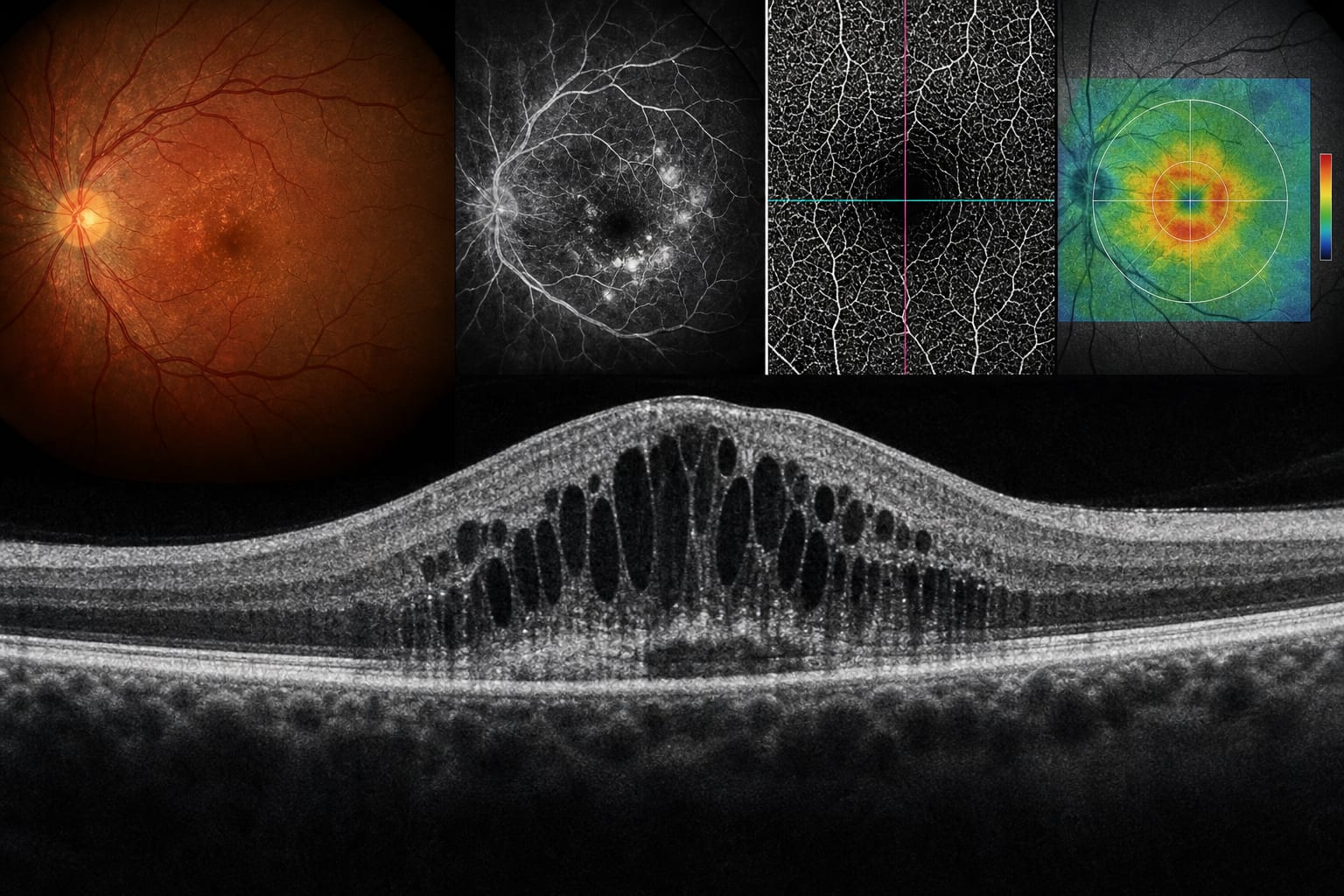
Multimodal imaging aids in characterizing retinal changes and identifying biomarkers.
DME can be classified into five clinical phenotypes based on imaging biomarkers.
Future advancements may enhance precision in DME phenotyping.
Guideline-Based Recommendations
Diagnosis
Utilize multimodal imaging techniques such as OCT, OCTA, and UWF-FA for comprehensive assessment.
Management
Implement phenotype-based classification to align therapeutic approaches with underlying mechanisms.
Monitoring & Follow-up
Regularly assess disease activity and visual prognosis using established imaging biomarkers.
Risks
Inadequate treatment responses may lead to ongoing visual impairment and increased healthcare costs.
Patient & Prescribing Data
Individuals with diabetic macular edema.
Consider alternative therapeutic options for patients with inadequate responses to standard anti-VEGF therapy.
Clinical Best Practices
Incorporate imaging biomarkers into treatment decision-making.
Adopt a dynamic optimization approach for therapy based on ongoing assessment of disease activity.