Intraoperative Evaluation of Sarcoma Surgical Margins With Indocyanine Green Fluorescence Imaging: Results From an Observational Study - Scorecard - MDSpire
Clinical Scorecard: Assessment of Surgical Margins in Sarcoma Procedures Using Indocyanine Green Fluorescence Imaging: Findings from an Observational Study
At a Glance
Category
Detail
Condition
Sarcoma
Key Mechanisms
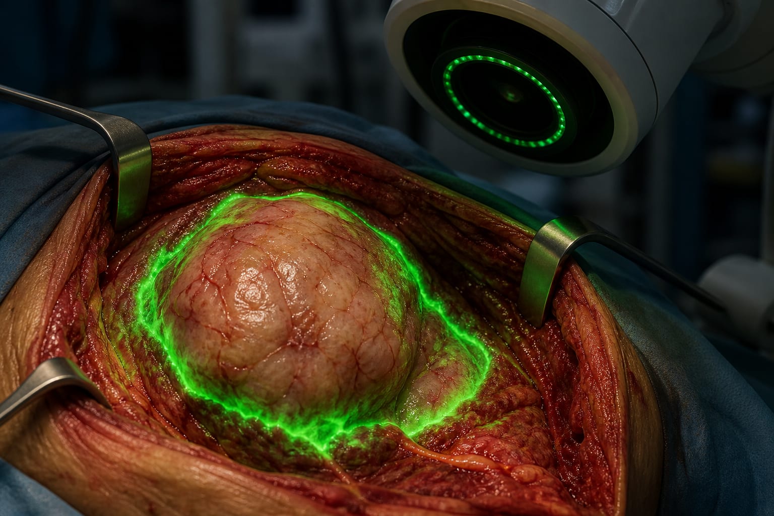
Indocyanine green (ICG) fluorescence imaging for assessing surgical margins.
Target Population
Adults and children with biopsy-confirmed soft tissue or bone sarcoma scheduled for margin-negative surgery.
Care Setting
Surgical oncology
Key Highlights
ICG fluorescence imaging shows high sensitivity in detecting positive margins in sarcoma surgery.
The study evaluated 51 patients, establishing a sarcoma-specific ICG protocol.
Positive surgical margins are linked to local recurrence and higher mortality.
Guideline-Based Recommendations
Diagnosis
Use ICG to characterize tumor margins during sarcoma surgery.
Management
Aim for negative histopathological margins (R0) during surgical resection.
Monitoring & Follow-up
Follow-up with clinical assessments and serial scans post-surgery.
Risks
Local recurrence rates of 10%-30% reported in sarcoma surgeries.
Patient & Prescribing Data
Patients aged 18 and older with confirmed soft tissue or bone sarcoma.
ICG administered intravenously prior to surgery to assess margins.
Clinical Best Practices
Utilize ICG imaging to enhance margin assessment during sarcoma surgeries.
Document all ICG findings and correlate with final pathology for accuracy.
by Sumail Bhogal, Matthew Gong, William Li, Brenda Diergaarde, Alma Heyl, Clark Roth, Lawrence Garvin, Alexander Hoffman, Amanda Locker, Ines Lohse, Maria Silvaggio, Stella Lee, Richard McGough, Kurt Weiss