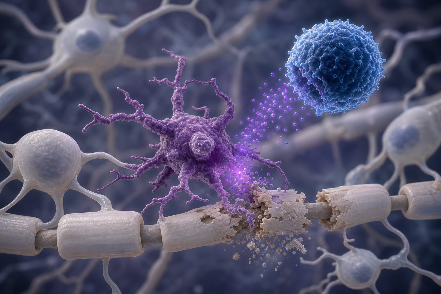
Clinical Scorecard: CXCL9 Correlates with Experimental Neuromyelitis Optica Spectrum Disorder Induced by Adoptive Transfer of Tfh and Th17 Cell Populations
At a Glance
Category
Detail
Condition
Neuromyelitis Optica Spectrum Disorder (NMOSD)
Key Mechanisms
Involvement of AQP4-specific Tfh and Th17 cells in neuroinflammation and demyelination.
Target Population
Patients with NMOSD, particularly those with AQP4 antibodies.
Care Setting
Experimental animal models and potential clinical implications.
Key Highlights
AQP4-reactive Tfh and Th17 cells induce significant neurological deficits in murine models.
CXCL9 is identified as a key chemokine associated with Tfh-mediated inflammation.
Histopathological analysis shows robust CNS immune cell infiltration and demyelination.
Guideline-Based Recommendations
Diagnosis
Detection of AQP4 antibodies in serum is a key diagnostic criterion for NMOSD.
Management
Adoptive transfer models can be used to study the pathogenesis of NMOSD.
Monitoring & Follow-up
Clinical assessment of neurological deficits and histopathological evaluation of CNS tissues.
Risks
Potential for severe neuroinflammation and demyelination associated with Tfh and Th17 cell activity.
Patient & Prescribing Data
Individuals diagnosed with NMOSD, particularly those with AQP4 antibodies.
Understanding Tfh and Th17 cell contributions may guide therapeutic strategies.
Clinical Best Practices
Utilize AQP4-knockout mouse models for studying NMOSD pathogenesis.
Incorporate flow cytometry for quantifying immune cell infiltration in CNS.