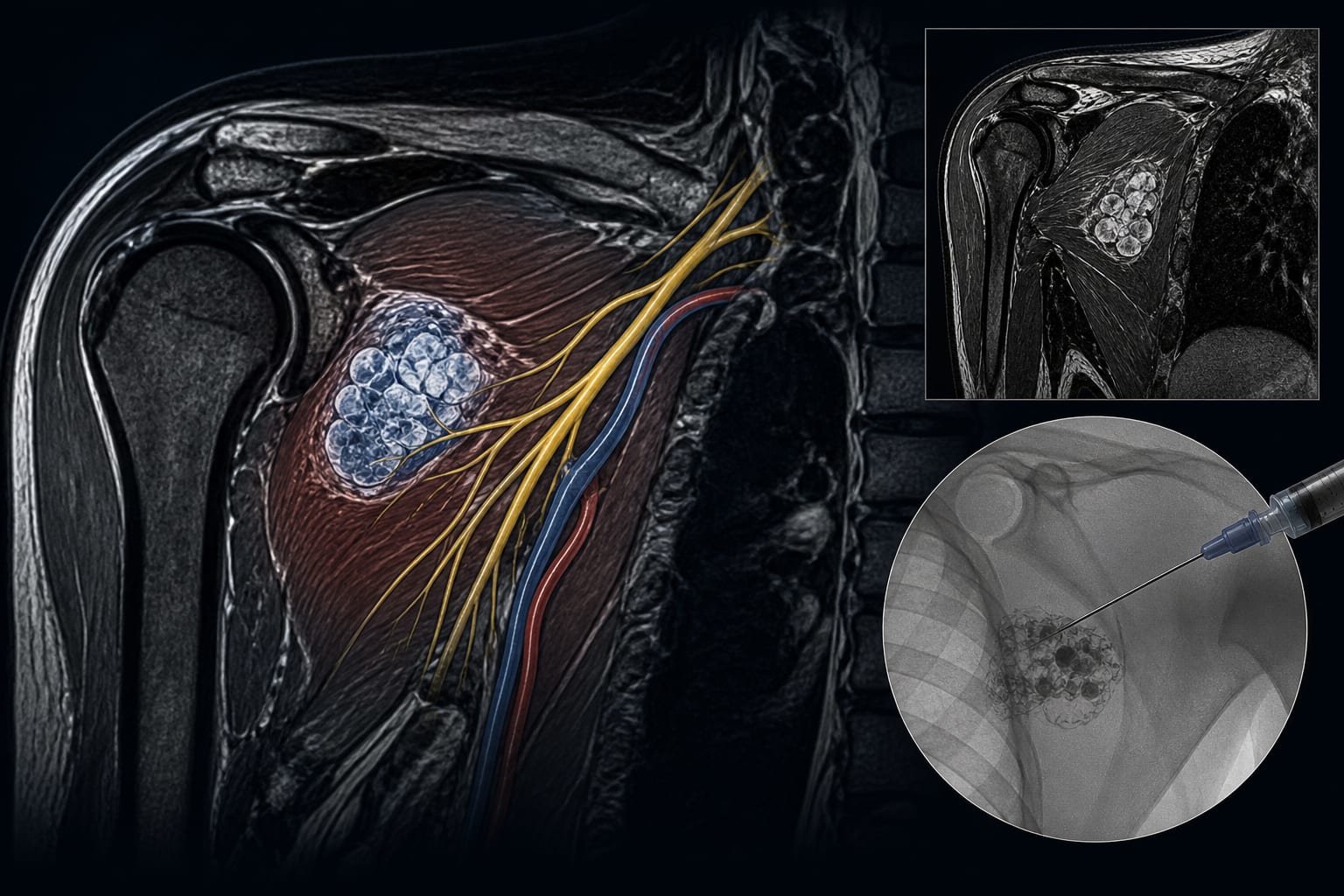
Uncommon Case of Intramuscular Venous Malformation in the Subscapularis Muscle Mimicking Traumatic Brachial Plexopathy
By
Jin Hang Loh
Zhun Shen Tan
Elaine Zi Fan Soh
Chia Hua Lim
Muhammad Shafiq Azhar
Jamari Sapuan
Shalimar Abdullah
September 9, 2026
Clinical Scorecard: Uncommon Case of Intramuscular Venous Malformation in the Subscapularis Muscle Mimicking Traumatic Brachial Plexopathy
At a Glance
Category Detail
Condition Intramuscular Venous Malformation Key Mechanisms Secondary compressive brachial plexopathy due to a venous malformation in the subscapularis muscle. Target Population Adults with vascular malformations causing neurological symptoms. Care Setting Specialized hand center
Key Highlights
Intramuscular venous malformation can mimic traumatic brachial plexopathy. MRI is crucial for diagnosing vascular malformations. Fluoroscopy-guided percutaneous bleomycin sclerotherapy was successfully used. Marked neurological recovery was observed post-treatment. Chronic compression from the lesion can lead to permanent neurological deficits. Guideline-Based Recommendations
Diagnosis
Use MRI to identify vascular lesions and assess their impact on surrounding structures. Management
Consider percutaneous sclerotherapy for symptomatic venous malformations. Monitoring & Follow-up
Ongoing surveillance of lesion size and neurological symptoms is recommended. Risks
Surgical resection carries a high risk of iatrogenic neurological injury. Patient & Prescribing Data
27-year-old male with no prior medical history.
Two sessions of percutaneous bleomycin sclerotherapy led to significant neurological improvement.
Clinical Best Practices
Multidisciplinary discussion is essential for treatment planning. Monitor motor and sensory function post-treatment to assess recovery. Related Resources & Content