Clinical Scorecard: Evaluating Flap Fixation Techniques to Reduce Seroma Development Post-Mastectomy: A Comprehensive Meta-Analysis
At a Glance
Category
Detail
Condition
Seroma formation following mastectomy
Key Mechanisms
Serous fluid collection under skin flaps or axillary dead space due to surgical dead space after mastectomy or axillary dissection
Target Population
Breast cancer patients undergoing mastectomy with or without axillary lymph node dissection
Care Setting
Surgical oncology and postoperative care settings
Key Highlights
Seroma incidence post-mastectomy varies widely from 3% to 90%, causing patient discomfort and potential complications.
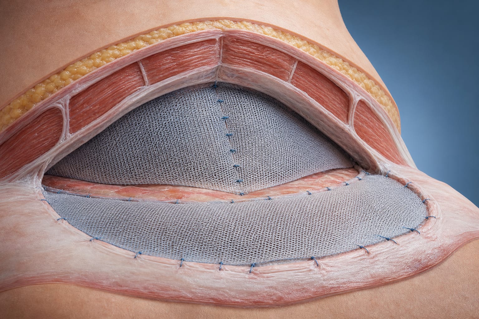
Flap fixation techniques, including quilting and tissue glue application, aim to reduce dead space and thus seroma formation.
Meta-analysis conducted using PRISMA guidelines and PICO framework to evaluate flap fixation versus no fixation on seroma and surgical site infection outcomes.
Employ flap fixation techniques such as quilting of skin flaps or adhesive tissue glue to reduce dead space and seroma formation.
Use closed-suction drainage as an adjunctive approach.
Monitoring & Follow-up
Monitor for seroma formation and surgical site infection postoperatively, especially in patients with risk factors like obesity and extensive axillary dissection.
Risks
Seroma can lead to repeated aspirations, infection risk, prolonged hospital stay, delayed wound healing, and delayed adjuvant therapy.
Patient & Prescribing Data
Breast cancer patients undergoing mastectomy with or without axillary lymph node dissection
Flap fixation reduces symptomatic seroma incidence compared to no fixation; demographic and oncological variables may influence outcomes.
Clinical Best Practices
Reduce dead space after mastectomy to minimize seroma formation using flap fixation techniques.
Perform thorough patient assessment including age, BMI, smoking status, tumor stage, and lymph node involvement to identify seroma risk.
Apply evidence-based surgical techniques and postoperative monitoring protocols to improve patient outcomes.
Use validated tools (Cochrane risk of bias, Newcastle–Ottawa Scale) to assess study quality when interpreting evidence.