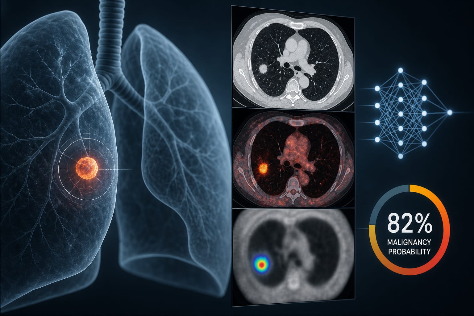
Clinical Scorecard: CT Features of Lung Cancer Lesions and Their Associations With Histologic Subtypes in Screening Programs
At a Glance
Category
Detail
Condition
Histologically verified lung cancers detected through low-dose CT screening, with radiologic features assessed by histologic subtype.
Key Mechanisms
The study evaluates associations between nodule diameter, volume, attenuation, location, and volume doubling time and lung cancer histology.
Target Population
NELSON screening participants aged 50–74 years who met high-risk smoking eligibility criteria.
Care Setting
Dutch lung cancer screening sites participating in the NELSON trial; retrospective observational analysis of screening and follow-up data.
Key Highlights
The study examined radiologic characteristics associated with histologic subtypes in histologically verified, screen-detected lung cancers.
Trained thoracic radiologists blinded to histology assessed lesion diameter, volume, attenuation, and location using standardized NELSON protocols.
The CT scan nearest histologic diagnosis was used to assess lesion features; volume doubling time was calculated from longitudinal imaging.
Two experienced thoracic radiologists retrospectively matched cancer diagnoses to index nodules using anatomical and morphological criteria, resolving discrepancies by consensus.
Guideline-Based Recommendations
Diagnosis
Histologic diagnoses were obtained from national pathology registries and assessed using the WHO classification of lung tumors.
Radiologic features were assessed on the CT scan nearest histologic diagnosis; VDT was derived from imaging at multiple time points.
Management
Monitoring & Follow-up
NELSON screening included baseline LDCT and three subsequent follow-up rounds.
Risks
The study population was restricted to participants meeting high-risk smoking eligibility criteria; findings may not describe other populations.
Nodule matching failures due to insufficient information about the cancer or nodule location led to exclusion.
Patient & Prescribing Data
Men and women aged 50–74 years who were current smokers or had quit within the preceding 10 years and met the trial's smoking-intensity and duration criteria.
This article excerpt describes an observational imaging study and does not provide treatment or prescribing recommendations.
Clinical Best Practices
Use standardized screening protocols and validated volumetric software when assessing nodule characteristics, as in the NELSON study.
When linking a cancer diagnosis to an index nodule retrospectively, apply predefined anatomical and morphological criteria and resolve disagreements by consensus.
Do not use PET/CT metabolic activity for lesion selection when it is not systematically available; it was not used in this study.
by Erick Suazo-Zepeda, Geertruida H. de Bock, Noa Antonissen, Colin Jacobs, Firdaus A. A. Mohamed Hoesein, Hester A. Gietema, Grigory Sidorenkov, Rozemarijn Vliegenthart, Marjolein A. Heuvelmans
The American Thoracic Society's 2026 International Conference brought together leading voices in pulmonary, critical care, and sleep medicine — and Temple Health was well represented.