Clinical Scorecard: Reassessing Surgical Interventions: Addressing Residual Tumors in Diffuse Glioma Patients through Second-look Surgery
At a Glance
Category
Detail
Condition
Diffuse glioma including glioblastoma (GBM)
Key Mechanisms
Surgical resection extent influences progression-free and overall survival; residual tumor volume impacts prognosis; early second-look surgery may address unexpected residual tumor
Target Population
Patients with diffuse glioma undergoing initial surgical resection, including those with residual tumor detected on early postoperative MRI
Care Setting
Neurosurgical and neuro-oncology referral centers with access to advanced imaging and multidisciplinary tumor boards
Key Highlights
Complete resection of contrast-enhancing tumor tissue improves progression-free survival in GBM.
Resection of non-contrast-enhancing tumor beyond residual volume < 5 cm³ further prolongs survival.
Early second-look surgery within 6 weeks can be considered for unexpected residual tumor based on resectability and patient status.
Guideline-Based Recommendations
Diagnosis
Use early postoperative MRI within 24–48 hours to assess extent of resection and detect residual tumor.
Employ gadolinium contrast-enhanced T1-weighted and T2/FLAIR sequences for volumetric tumor assessment.
Classify resections in GBM according to RANO resect classification system for prognostic evaluation.
Management
Consider early second-look surgery within 6 weeks for unexpected residual tumor after initial resection.
Decision for re-resection should be multidisciplinary, considering tumor location, eloquence, patient performance status, and surgical safety.
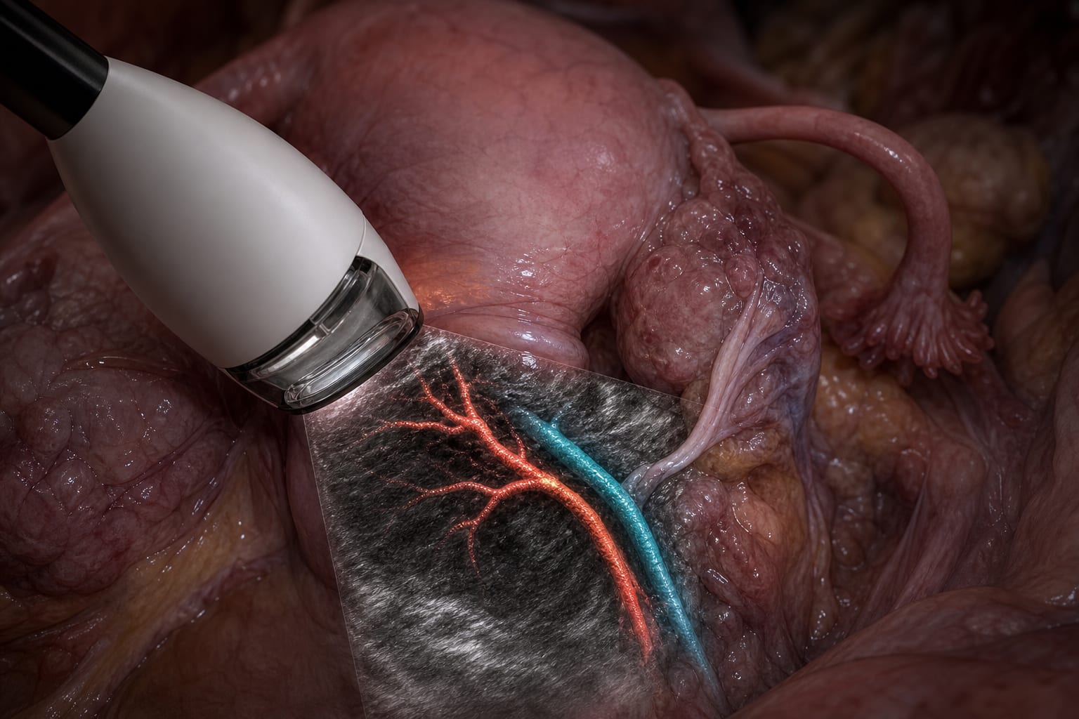
Utilize intraoperative adjuncts such as neuronavigation, ioUS, 5-ALA fluorescence, and ioMRI to optimize tumor margin delineation.
Monitoring & Follow-up
Assess functional status using Karnofsky Performance Score (KPS) and neurological status with NIH Stroke Scale (NIHSS).
Monitor progression-free survival and overall survival using radiological progression and mortality data.
Perform postoperative MRI volumetry to guide further treatment decisions.
Risks
Potential neurological deficits from aggressive or supramaximal resections, especially in eloquent brain areas.
Surgical risks associated with re-operation including infection, hemorrhage, and anesthesia-related complications.
Balancing extent of resection with preservation of neurological function is critical.
Patient & Prescribing Data
Diffuse glioma patients undergoing initial and possible second-look surgical resections.
Early second-look surgery is selectively applied based on residual tumor volume, resectability, and patient condition, potentially improving survival outcomes without predefined volumetric thresholds.
Clinical Best Practices
Perform early postoperative MRI within 24–48 hours to accurately assess residual tumor volume.
Use multidisciplinary tumor board discussions to guide decisions on early second-look surgery.
Incorporate advanced intraoperative imaging and neurophysiological monitoring to maximize safe extent of resection.
Prioritize preservation of neurological function, especially in patients with longer expected survival.
Apply standardized volumetric tools and RANO classification to objectively evaluate extent of resection.
Severe social jet lag among surgeons was associated with higher rates of major adverse events, independent of sleep duration, workload, and patient risk.
These 10 states make it more practical for physicians to participate in hospital ownership by aligning statutory structure, corporate practice of medicine rules, and population trends.