Case Report: Retained gallstone migrating to the chest wall after cholecystectomy presenting as chronic abscess, sinus tract, and inflammatory pseudotumor - Scorecard - MDSpire
Advertisement
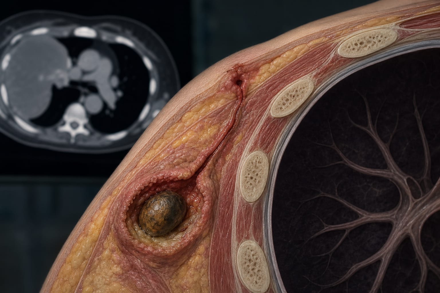
Case Study: Migration of a Retained Gallstone to the Chest Wall Following Cholecystectomy Resulting in Chronic Abscess, Sinus Tract, and Inflammatory Pseudotumor
Clinical Scorecard: Case Study: Migration of a Retained Gallstone to the Chest Wall Following Cholecystectomy Resulting in Chronic Abscess, Sinus Tract, and Inflammatory Pseudotumor
At a Glance
Category
Detail
Condition
Retained Gallstone Migration
Key Mechanisms
Gallstone spillage during laparoscopic cholecystectomy leading to chronic inflammation and abscess formation.
Target Population
Patients with a history of laparoscopic cholecystectomy.
Care Setting
Post-surgical follow-up and management of complications.
Key Highlights
Gallbladder perforation and stone spillage occur in 10%-40% and 6%-10% of laparoscopic cholecystectomy cases, respectively.
Retained gallstones can migrate to extra-abdominal sites, causing chronic pathology.
Imaging findings may mimic malignancy, necessitating careful evaluation.
Complete surgical excision of the retained stone is curative.
Guideline-Based Recommendations
Diagnosis
Use CT and MRI to evaluate suspected retained gallstones in post-cholecystectomy patients.
Management
Surgical excision of the retained gallstone and associated abscess.
Monitoring & Follow-up
Follow-up imaging to assess for recurrence or complications.
Risks
Potential for chronic abscess, sinus tract formation, and misdiagnosis as malignancy.
Patient & Prescribing Data
Post-cholecystectomy patients presenting with unexplained chest wall pathology.
Surgical intervention is necessary for symptomatic retained gallstones.
Clinical Best Practices
Maintain a high index of suspicion for retained gallstones in patients with a history of cholecystectomy presenting with unusual symptoms.
Utilize imaging modalities effectively to differentiate between benign and malignant lesions.
An ancillary analysis found less than 2-percentage-point differences in 10-year recurrence across margin thresholds, with no statistically significant adjusted association.