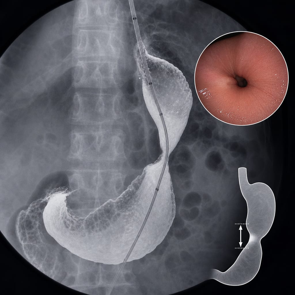
Fluoroscopic Metrics for Diagnosing and Quantifying Gastric Sleeve Stenosis
-
By
-
Samuel Tanner
-
Susie Min
-
Jessica X. Yu
-
Sarah Volk
-
Allison R. Schulman
-
May 5, 2026
Clinical Scorecard: Fluoroscopic Metrics for Diagnosing and Quantifying Gastric Sleeve Stenosis
At a Glance
| Category | Detail |
|---|
| Condition | Gastric Sleeve Stenosis (GSS) |
| Key Mechanisms | Obstructive symptoms due to anatomic narrowing or twisting of the gastric sleeve. |
| Target Population | Patients who have undergone laparoscopic sleeve gastrectomy. |
| Care Setting | Endoscopy centers performing bariatric procedures. |
Key Highlights
- GSS occurs in 1-4% of patients post-sleeve gastrectomy.
- Symptoms include regurgitation, dyspepsia, and dysphagia.
- Diagnosis can be challenging due to non-specific symptoms and lack of objective data.
- Fluoroscopy during endoscopy can provide objective metrics for GSS severity.
- Impedance planimetry can measure luminal diameter and distensibility.
Guideline-Based Recommendations
Diagnosis
- Upper endoscopy is recommended for reliable identification of luminal narrowing.
- Fluoroscopy can be used to assess the presence and severity of GSS.
Management
- Pneumatic dilation may be performed under endoscopic and fluoroscopic guidance.
Monitoring & Follow-up
- Regular assessment of symptoms and endoscopic evaluation for patients with persistent symptoms.
Risks
- Potential complications from endoscopic procedures and pneumatic dilation.
Patient & Prescribing Data
Patients with symptoms suggestive of gastric sleeve stenosis post-sleeve gastrectomy.
Conservative management for early post-operative GSS; further evaluation required for persistent symptoms.
Clinical Best Practices
- Utilize fluoroscopic metrics to enhance diagnostic accuracy for GSS.
- Ensure experienced endoscopists perform evaluations to minimize diagnostic errors.
References