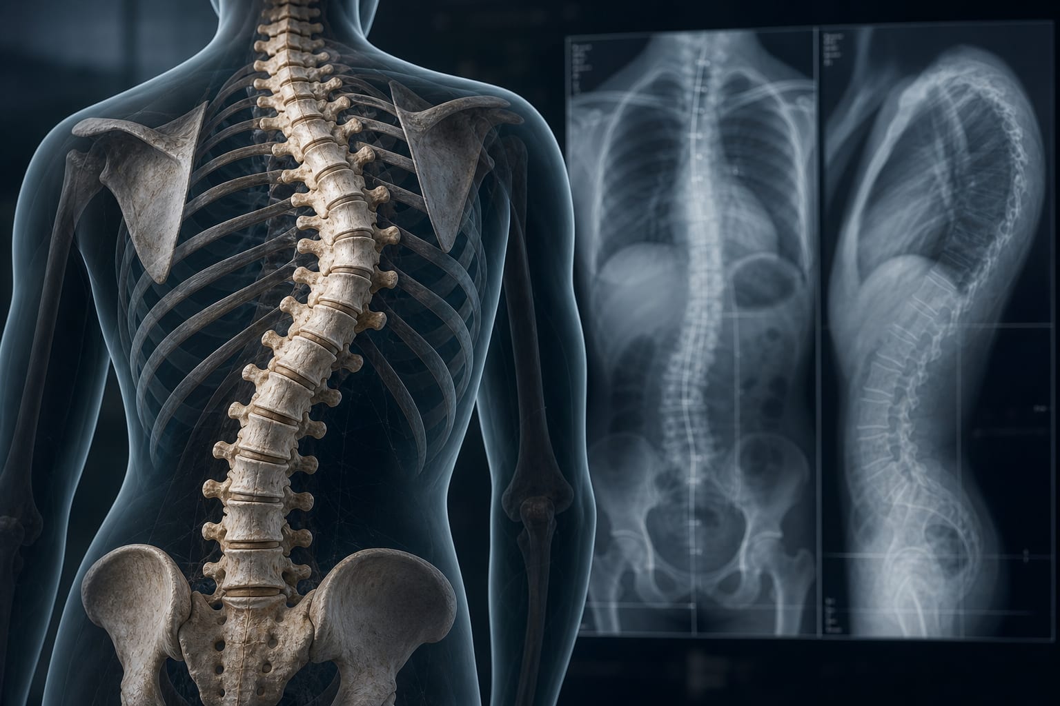
Biomechanics of chest wall injury: implications for surgical stabilization and failure prevention
-
By
-
Vladislav Muldiiarov
-
Zachary M. Bauman
-
July 7, 2026
-
Clinical Scorecard: Chest Wall Injury Biomechanics: Considerations for Surgical Stabilization and Prevention of Complications
At a Glance
| Category | Detail |
|---|
| Condition | Chest wall injury |
| Key Mechanisms | Impairment of ventilation due to pain and loss of thoracic cage stability. |
| Target Population | Patients with rib fractures, particularly older adults and those with multiple rib fractures. |
| Care Setting | Acute care surgery |
Key Highlights
- Surgical stabilization of rib fractures (SSRF) reduces ventilator days and pulmonary complications.
- Variability in operative technique and construct design exists in SSRF.
- Common modes of fixation failure can be linked to inadequate matching of implant behavior to physiologic loading.
- Thoracic cage biomechanics should inform surgical planning to reduce complications.
- Delayed deterioration in patients with chest wall injury can occur 48-72 hours post-injury.
Guideline-Based Recommendations
Diagnosis
- Assess chest wall injury through imaging and clinical evaluation.
Management
- Utilize SSRF for selected patients with multiple severely displaced rib fractures.
Monitoring & Follow-up
- Monitor for complications such as atelectasis, pneumonia, and respiratory failure.
Risks
- Inadequate fixation can lead to hardware-related symptoms, nonunion, or construct failure.
Patient & Prescribing Data
Adults with chest wall trauma, particularly those with multiple rib fractures.
Surgical intervention should be tailored based on specific instability patterns and functional impairment.
Clinical Best Practices
- Incorporate biomechanical principles into surgical planning for chest wall injuries.
- Select fixation constructs that match the physiologic loading conditions of the thoracic cage.
Related Resources & Content