Clinical Scorecard: Prenatal MRI Assessment of Fetuses with Congenital Cytomegalovirus Infection: A Comparison of Valacyclovir Treatment and No Treatment
At a Glance
Category
Detail
Condition
Congenital Cytomegalovirus Infection
Key Mechanisms
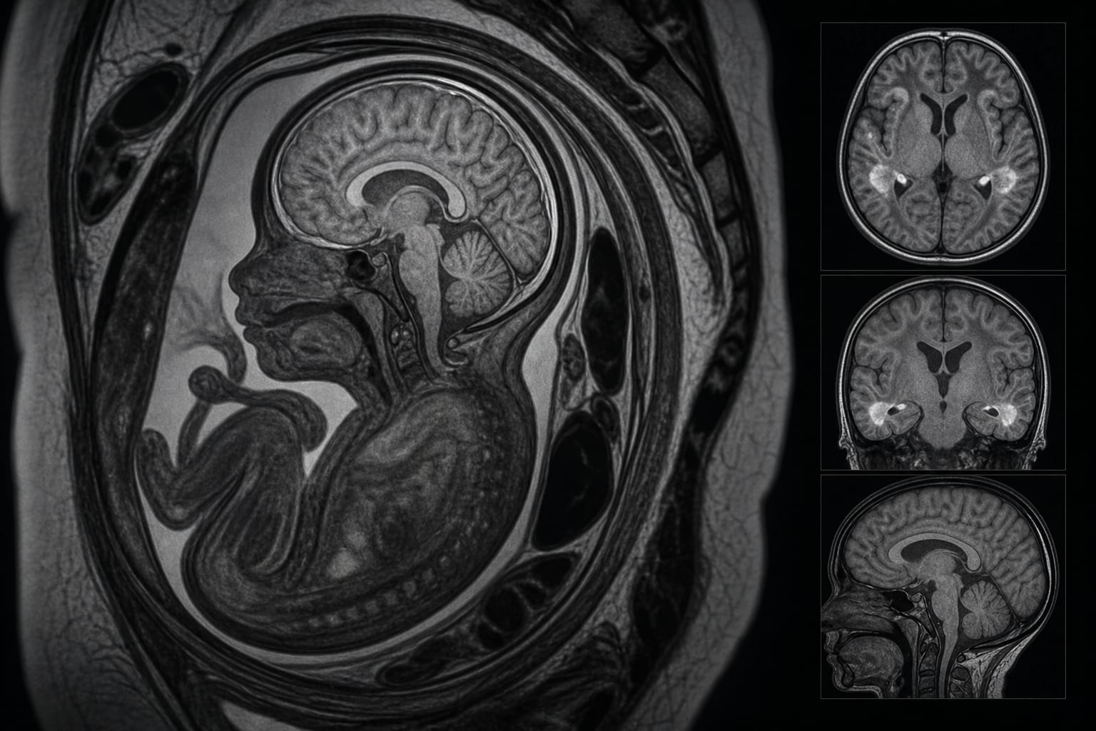
Fetal MRI detects brain abnormalities associated with cCMV infection.
Target Population
Fetuses with confirmed congenital CMV infection.
Care Setting
Prenatal imaging and assessment.
Key Highlights
cCMV is the leading cause of non-genetic sensorineural hearing loss and neurodevelopmental impairment.
Fetal MRI provides superior contrast resolution compared to ultrasound.
Oral valacyclovir may reduce viral replication and improve neurological outcomes.
The study focuses on the impact of valacyclovir on 'minor' brain lesions.
Fetuses with major brain abnormalities were excluded from the study.
Guideline-Based Recommendations
Diagnosis
Confirm maternal CMV infection via seroconversion.
Confirm fetal infection through PCR or viral isolation from amniotic fluid.
Management
Consider oral valacyclovir (8 g/day) for maternal treatment.
Monitoring & Follow-up
Conduct serial fetal MRI examinations to assess lesion progression.
Risks
Up to 10-15% of infected fetuses may develop long-term sequelae.
Patient & Prescribing Data
Fetuses with confirmed cCMV infection and no major brain anomalies.
Valacyclovir treatment may influence the development of subtle MRI abnormalities.
Clinical Best Practices
Utilize fetal MRI for detailed assessment of brain abnormalities in cCMV.
Select cases for treatment based on the absence of severe brain anomalies.
The partner in the next room, the hormone in the blister pack, the cat on the couch, the minute-long chair stand. Several new studies suggest the factor shaping outcomes may be the one clinicians aren’t routinely measuring.