Retinal Imaging Model Aids CAD Detection
Fundus-based model showed moderate-to-good accuracy and improved performance when combined with clinical risk factors in a high-risk cohort
By
Andrea Surnit
April 17, 2026
Clinical Scorecard: Retinal Imaging Model Aids CAD Detection
At a Glance
Category Detail
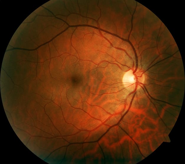
Condition Coronary Artery Disease (CAD) Key Mechanisms Quantitative retinal vascular parameters reflecting systemic vascular health and coronary vessel pathology Target Population Patients with suspected angina undergoing coronary angiography Care Setting Cardiology diagnostic evaluation, outpatient or hospital-based angiography centers
Key Highlights
Combined retinal imaging and clinical risk factor model achieved AUROC of 0.802 for CAD detection. Retinal variables independently associated with CAD included decreased fractal dimension, reduced optic disc axis ratio, and shorter optic disc-to-macula distance. Retinal imaging model showed higher sensitivity (0.797) than specificity (0.679), suggesting utility in identifying patients at increased CAD risk. Guideline-Based Recommendations
Diagnosis
Consider retinal vascular phenotyping as an adjunct to clinical risk assessment for CAD detection. Use non-mydriatic fundus photography within 24 hours prior to coronary angiography for retinal imaging. Management
Incorporate retinal imaging findings with established clinical risk factors such as sex, dyslipidemia, and lipid profiles to stratify CAD risk. Monitoring & Follow-up
Monitor retinal vascular parameters alongside traditional cardiovascular risk markers to evaluate disease progression or risk. Risks
Recognize limitations due to retrospective, single-center design and high-risk study population limiting generalizability. Be cautious interpreting retinal imaging results in populations differing from the predominantly Han Chinese cohort studied. Patient & Prescribing Data
High-risk patients referred for first-time coronary angiography with suspected angina
Retinal imaging combined with clinical risk factors may improve CAD risk stratification but requires further validation before routine clinical use.
Clinical Best Practices
Use explicitly quantified retinal vascular features to improve interpretability over black-box deep learning models. Employ non-mydriatic fundus photography to facilitate integration into routine clinical workflows. Combine retinal imaging data with traditional clinical risk factors for enhanced CAD detection accuracy. Interpret retinal imaging results within the context of patient demographics and clinical risk profile. Related Resources & Content