Label-Free Multiphoton Microscopy for intraoperative Identification of Glioma Features and Tumor Heterogeneity
-
By
-
Duncker, León V.
-
Richter, Sven
-
Galli, Roberta
-
Meinhardt, Matthias
-
Hoffmann, Leon C.
-
Kirsche, Katrin
-
Großer, Lidia
-
Temme, Achim
-
Eyüpoglu, Ilker Y.
-
Uckermann, Ortrud
-
June 22, 2026
-
Clinical Scorecard: Intraoperative Characterization of Glioma Characteristics and Tumor Diversity Using Label-Free Multiphoton Microscopy
At a Glance
| Category | Detail |
|---|
| Condition | Diffuse gliomas |
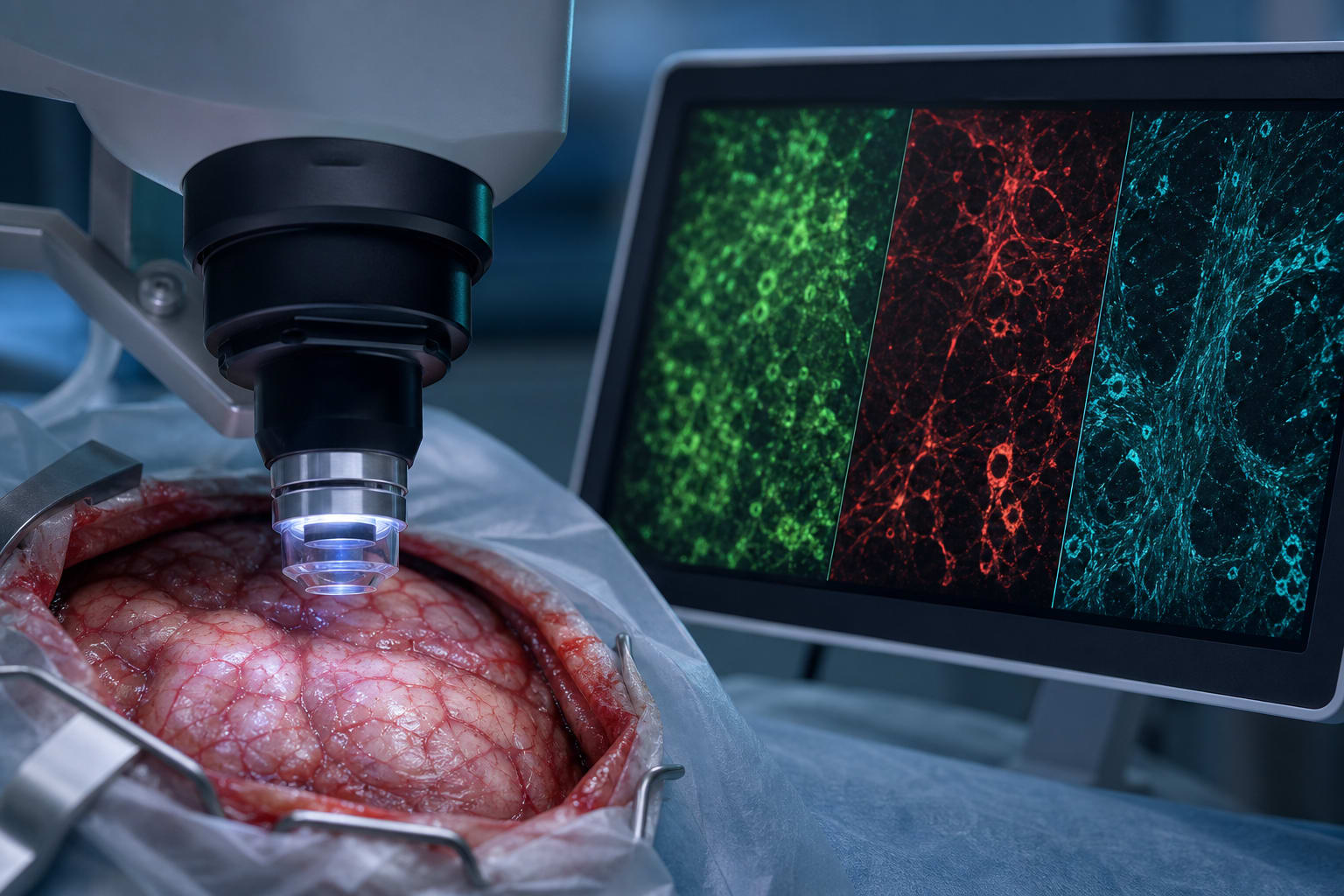
| Key Mechanisms | Label-free multiphoton imaging modalities including CARS, AF, and SHG for tissue characterization |
| Target Population | Patients with gliomas including glioblastoma, astrocytoma, and oligodendroglioma |
| Care Setting | Intraoperative surgical environment |
Key Highlights
- Intraoperative imaging can adapt surgical strategy based on tumor subtype and grade.
- CARS detected lipid droplets, AF showed cellular structures, and SHG visualized blood vessels.
- Statistically significant differences in feature prevalence among tumor types.
- Numerical scoring system based on tissue features indicative of malignancy.
- Highest scores observed in WHO grade 4 tumors.
Guideline-Based Recommendations
Diagnosis
- Utilize multimodal imaging for intraoperative glioma grading.
Management
- Adapt surgical strategy based on intraoperative tumor characterization.
Monitoring & Follow-up
- Evaluate tumor features during surgery for real-time decision making.
Risks
- Consider variability in tissue feature prevalence among patients.
Patient & Prescribing Data
102 glioma patients across various grades.
Intraoperative imaging provides enhanced information on tumor malignancy.
Clinical Best Practices
- Incorporate label-free multiphoton imaging in surgical protocols for gliomas.
- Train surgical teams on interpreting imaging features for real-time decision making.
Related Resources & Content