Correlation of histologic, imaging, and artificial intelligence features in NAFLD patients, derived from Gd-EOB-DTPA-enhanced MRI: a proof-of-concept study - Scorecard - MDSpire
Advertisement
Correlation of histologic, imaging, and artificial intelligence features in NAFLD patients, derived from Gd-EOB-DTPA-enhanced MRI: a proof-of-concept study
Clinical Scorecard: Association of Histological, Imaging, and AI Characteristics in Patients with NAFLD Using Gd-EOB-DTPA-Enhanced MRI: A Proof-of-Concept Investigation
At a Glance
Category
Detail
Condition
Non-alcoholic fatty liver disease (NAFLD), including simple steatosis and non-alcoholic steatohepatitis (NASH)
Key Mechanisms
Liver fat accumulation, inflammation, fibrosis, and hepatocyte injury leading to progressive hepatic dysfunction
Target Population
Patients with clinical suspicion of fatty liver disease and elevated liver enzymes
Care Setting
Tertiary academic institution with access to advanced MRI and histopathology
Key Highlights
NASH diagnosis currently relies on invasive liver biopsy with limitations including bleeding risk and sampling errors.
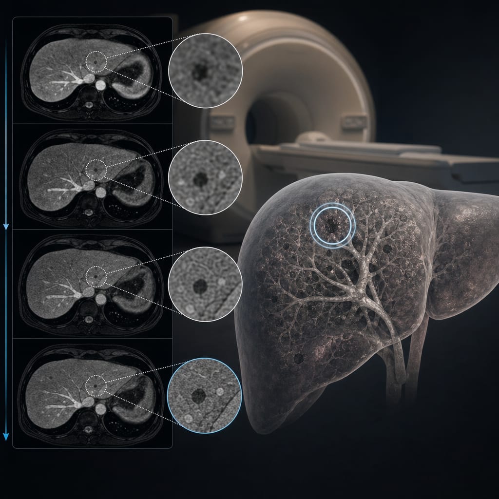
Gd-EOB-DTPA-enhanced MRI combined with chemical shift imaging (CSI) and AI-based texture analysis can non-invasively differentiate NASH from simple steatosis.
A hybrid unsupervised and supervised deep learning approach (UDC) applied to MRI data shows promise in identifying predictive imaging patterns correlating with histopathology.
Guideline-Based Recommendations
Diagnosis
Use liver biopsy as the gold standard for differentiating simple steatosis from NASH.
Consider multiparametric MRI techniques including PDFF quantification and MR elastography for non-invasive assessment.
Employ Gd-EOB-DTPA-enhanced MRI with relative liver enhancement (RLE) and fat fraction (FF) measurements to improve diagnostic accuracy.
Integrate AI-based imaging analysis to enhance differentiation between NASH and simple steatosis.
Management
Lifestyle modifications remain first-line for simple steatosis.
Pharmacotherapy may be required for NASH to prevent progression to cirrhosis or hepatocellular carcinoma.
Early diagnosis and intervention are critical to mitigate end-stage liver disease complications.
Monitoring & Follow-up
Non-invasive imaging modalities such as Gd-EOB-DTPA-enhanced MRI can be used for longitudinal monitoring.
Serum biomarkers and fibrosis scores (FIB-4, NFS, ALBI, APRI) should be measured alongside imaging for risk stratification.
Risks
Liver biopsy carries bleeding risk, especially in patients with coagulopathy.
Conventional imaging and serum markers lack specificity to reliably distinguish NASH from simple steatosis.
High prevalence of NAFLD limits feasibility of universal biopsy.
Patient & Prescribing Data
Adults with histologically confirmed simple steatosis or NASH undergoing Gd-EOB-DTPA-enhanced MRI
Non-invasive imaging combined with AI may guide clinical decision-making and reduce reliance on invasive biopsy for diagnosis and monitoring.
Clinical Best Practices
Obtain informed consent and adhere to ethical protocols for imaging and biopsy procedures.
Exclude patients with confounding liver diseases and significant alcohol intake per guidelines.
Use standardized MRI protocols including CSI and hepatobiliary phase imaging with Gd-EOB-DTPA.
Apply validated histopathological scoring systems (SAF) for biopsy interpretation.
Incorporate AI-driven texture analysis to enhance diagnostic accuracy from imaging data.
Combine imaging findings with serum biomarkers and fibrosis scores for comprehensive assessment.
by Nina Bastati, Matthias Perkonigg, Daniel Sobotka, Sarah Poetter-Lang, Romana Fragner, Andrea Beer, Alina Messner, Martin Watzenboeck, Svitlana Pochepnia, Jakob Kittinger, Alexander Herold, Antonia Kristic, Jacqueline C. Hodge, Stefan Traussnig, Michael Trauner, Ahmed Ba-Ssalamah, Georg Langs