Neurosurgery and the glymphatic system
-
By
-
Per Kristian Eide
-
June 21, 2024
-
0 min
Clinical Scorecard: The Role of the Glymphatic System in Neurosurgery
At a Glance
| Category | Detail |
|---|---|
| Condition | Glymphatic system dysfunction and its implications in CNS diseases |
| Key Mechanisms | Brain-wide perivascular transport pathway clearing metabolic waste and facilitating substance transport |
| Target Population | Patients with CNS diseases including dementia, stroke, traumatic brain injury, and systemic diseases affecting glymphatic function |
| Care Setting | Neurosurgical and neurological clinical settings with advanced imaging capabilities |
Key Highlights
- The glymphatic system is most active during sleep and declines with aging and systemic diseases like hypertension and diabetes.
- Impaired glymphatic function is linked to accumulation of toxic proteins (amyloid-β, tau, α-synuclein) implicated in Alzheimer’s and Parkinson’s diseases.
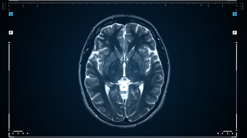
- Intrathecal contrast-enhanced MRI is currently considered the gold standard for assessing glymphatic function in humans despite requiring spinal puncture.
Guideline-Based Recommendations
Diagnosis
- Use intrathecal gadobutrol-enhanced MRI to visualize glymphatic transport in humans.
- Consider non-invasive MRI techniques such as DTI-ALPS, enlarged perivascular spaces assessment, and magnetic resonance encephalography (MREG) with caution due to limitations.
Management
- Recognize the potential role of glymphatic dysfunction in neurosurgical diseases including brain edema post-stroke and traumatic brain injury.
- Monitor and address systemic conditions like hypertension and diabetes that may impair glymphatic function.
Monitoring & Follow-up
- Employ imaging modalities to evaluate glymphatic function changes in response to interventions when feasible.
- Be aware of the current scarcity and limitations of clinical methods for glymphatic assessment.
Risks
- Intrathecal administration of gadobutrol is off-label and requires spinal puncture, with theoretical concerns about toxicity and brain deposition, though no adverse effects have been observed in hundreds of patients.
- Intravenous contrast-enhanced MRI may confound glymphatic and vascular tracer signals due to blood-brain barrier leakage.
Patient & Prescribing Data
Patients undergoing evaluation for CNS diseases or neurosurgical conditions where glymphatic function assessment is relevant
Intrathecal gadobutrol doses of 0.25 to 0.50 mmol have been used safely for glymphatic imaging; intravenous gadobutrol doses are higher but less specific for glymphatic assessment.
Clinical Best Practices
- Prefer intrathecal contrast-enhanced MRI for direct visualization of glymphatic transport despite procedural invasiveness.
- Interpret non-invasive imaging methods (DTI-ALPS, PVS burden, MREG) cautiously due to their indirect measures and potential confounders.
- Consider patient age and systemic disease status when evaluating glymphatic function as these factors influence efficacy.
- Remain updated on emerging methodologies and evidence regarding human glymphatic system function and its clinical relevance.
References
- Iliff et al., 2012 - Discovery of the glymphatic system
- Intrathecal gadobutrol MRI studies
- DTI-ALPS technique and limitations
- Glymphatic dysfunction in dementia and CNS diseases
- Magnetic resonance encephalography (MREG) for brain pulsations
This content is an AI-generated, fully rewritten summary based on a published scholarly article. It does not reproduce the original text and is not a substitute for the original publication. Readers are encouraged to consult the source for full context, data, and methodology.