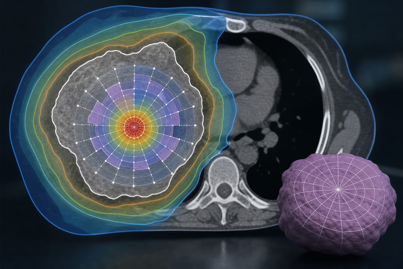
Clinical Scorecard: Extracellular Volume from MRI as an Indicator of Cardiotoxicity in Cancer Treatment: A Systematic Review and Meta-Analysis
At a Glance
Category
Detail
Condition
Cancer therapy-related cardiotoxicity
Key Mechanisms
Cardiomyocyte death leading to myocardial edema and fibrosis, detectable by increased myocardial extracellular volume (ECV) via MRI T1 mapping
Target Population
Patients undergoing cardiotoxic cancer therapies such as anthracyclines, chest radiotherapy, monoclonal antibodies, and small molecule inhibitors
Care Setting
Cardio-oncology clinical settings with access to cardiac MRI and echocardiography
Key Highlights
Cancer therapy-related cardiac dysfunction is defined by a ≥10% decline in left ventricular ejection fraction (LVEF).
Conventional biomarkers and echocardiography may not detect early myocardial damage due to cardiac functional reserve.
MRI-derived extracellular volume (ECV) estimation via T1 mapping is a promising biomarker for early detection of myocardial inflammation and fibrosis associated with cardiotoxicity.
Guideline-Based Recommendations
Diagnosis
Use echocardiography to assess LVEF and myocardial strain for detection of cancer therapy-related cardiotoxicity.
Monitor relevant serum biomarkers as adjunctive tools.
Consider cardiac MRI with T1 mapping to estimate myocardial ECV for early detection of subclinical myocardial changes.
Management
Adjust cancer therapy regimens upon detection of subclinical cardiotoxicity, e.g., pre-treatment with dexrazoxane in anthracycline-based chemotherapy.
Implement personalized follow-up schemes based on early biomarker detection.
Monitoring & Follow-up
Perform serial echocardiographic assessments of LVEF and strain during and after cancer therapy.
Use MRI-derived ECV measurements to monitor myocardial tissue changes over time.
Risks
Delayed detection of cardiotoxicity may lead to irreversible myocardial damage and increased morbidity and mortality.
Lack of sensitive biomarkers may result in overt heart failure before intervention.
Patient & Prescribing Data
Patients receiving cardiotoxic cancer therapies, particularly anthracycline-based regimens
Early detection of increased myocardial ECV can guide initiation of cardioprotective treatments such as dexrazoxane and tailored monitoring to prevent progression to overt cardiotoxicity.
Clinical Best Practices
Incorporate cardiac MRI with T1 mapping for ECV estimation in cardio-oncology protocols to detect early myocardial changes.
Use a multidisciplinary approach combining imaging, biomarkers, and clinical assessment for comprehensive cardiotoxicity surveillance.
Exclude patients with pre-existing cardiac comorbidities when interpreting ECV values to avoid confounding.
Apply standardized MRI acquisition protocols and timing (≥3 months post-therapy) for consistent ECV measurement.