Cross-software comparison shows strong agreement for quantitative indocyanine green fluorescence angiography in reconstructive surgery - Scorecard - MDSpire

Cross-software comparison shows strong agreement for quantitative indocyanine green fluorescence angiography in reconstructive surgery

  • By

  • Guy Oster

  • Lasse W. P. van ‘t Hof

  • Daniel M. de Bruin

  • Caroline Driessen

  • June 24, 2026

  • 0 min

Share

Clinical Scorecard: Evaluation of Consistency in Quantitative Indocyanine Green Fluorescence Angiography Across Different Software Platforms in Reconstructive Surgery

At a Glance

CategoryDetail
ConditionQuantitative Indocyanine Green Fluorescence Angiography
Key Mechanisms
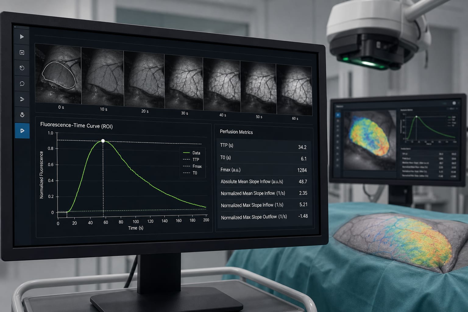
Assessment of intraoperative tissue perfusion using fluorescence-time curves derived from indocyanine green injections.
Target PopulationPatients undergoing reconstructive surgery.
Care SettingIntraoperative assessment in surgical settings.

Key Highlights

  • Excellent agreement for time-to-peak (TTP) with ICC = 0.979.
  • Normalized mean slope inflow showed good agreement (ICC = 0.944).
  • Significant systematic differences observed for six out of seven parameters.
  • AMS platform reported higher TTP values compared to EPA (p < 0.001).
  • Bland–Altman analysis indicated wide limits of agreement for TTP.

Guideline-Based Recommendations

Diagnosis

  • Use quantitative indocyanine green fluorescence angiography for objective perfusion assessment.

Management

  • Consider TTP and normalized mean slope inflow as reliable metrics for perfusion thresholds.

Monitoring & Follow-up

  • Evaluate consistency of Q-ICG-FA parameters across different software platforms.

Risks

  • Potential for interobserver variability in qualitative assessments of perfusion.

Patient & Prescribing Data

Patients undergoing various types of reconstructive surgery.

ICG administered intravenously at a dose of 0.1 mg/kg for perfusion assessment.

Clinical Best Practices

  • Standardize imaging protocols for Q-ICG-FA to enhance reproducibility.
  • Utilize both AMS and EPA software for comparative analysis of perfusion parameters.

Related Resources & Content

Original Source(s)

Related Content